

Pathologist.. and I have a dream! Founder @amontasserlab
Egypt
Joined December 2011
- Tweets2.4K
- Following764
- Followers2K
- Likes3.3K




Pinned Tweet
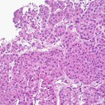
Surgeon: it looks like GIST but the endoscopic biopsy report says adenocarcinoma!
Me: well.. I’ve got news 😅
#pathology #surprise #GIpath #PathTwitter #collision #Tumor


- AI is gonna replace pathologists!
- Pathologists with AI:
🤖 Made with AI

ALT Mucinous adenocarcinoma with signet ting cells

Reason N 32584 I don’t do ovary frozen sections 😅
Great illustration 👌🏽

UGLY NUCLEI. BEAUTIFUL CLEAR CELLS. A TERRIFYING DIAGNOSTIC TRAP! 🔬
A 31-year-old woman. 27 weeks pregnant. A massive 12.5 cm ovarian cyst.
Under the microscope?
Bizarre, pleomorphic nuclei. Striking clear-cell cytoplasm. Cells that could make you fear ovarian clear cell carcinoma!
But the diagnosis?
Large solitary luteinized follicle cyst of pregnancy.
Completely BENIGN!
Pregnancy-related hormonal changes can produce astonishing cellular appearances that mimic malignancy.
A wrong diagnosis could mean unnecessary radical surgery and devastating consequences for a young mother.
The lesson?
Never let ugly nuclei dictate the diagnosis.
Never interpret high-power morphology without low-power architecture.
And NEVER underestimate clinical history.
Sometimes the most frightening cells are completely innocent.
#Pathology #WomensHealth #Pregnancy #DiagnosticPitfalls


Ahmed Montasser retweeted

When can HER2 IHC help in DCIS?
In an intraductal proliferation with apocrine features and intermediate- to high-grade atypia
Diffuse strong score 3+ HER2 staining supports DCIS and is not expected in hyperplasia
HER2 does not help with low-grade DCIS, or with ADH vs borderline DCIS
Dr. Allison. 2026 Diagnostic Pathology Update #USCAP #pathology #pathX


🔬 The triad of celiac disease:
1️⃣ Intraepithelial lymphocytosis
2️⃣ Crypt hyperplasia
3️⃣ Villous atrophy
#Pathology #GIPath #PathX #PathTwitter


F 80+ Y 👵🏽 Breast mass
🔬 APOCRINE CARCINOMA
TNBC
AR1 ➕ (shown)
#BreastCancerAwareness #Pathology #PathX #PathTwitter #PinkOctober


📍Trapani, Sicily 🇮🇹
14 hours travel 🧳
2 planes ✈️ ✈️ and a bus 🚍
100% worth it!!
#GUpath #Pathology #Education




Ahmed Montasser retweeted


Answer:
✅Cryoablation treatment effect for prostatic adenocarcinoma
-Can cause this grayish, degenerative, edematous, or fibrotic appearance of the prostatic stroma with a paucity of glands.
-No residual adenocarcinoma in this field.
#Pathology #PathX #PathTwitter
Ahmed Montasser retweeted

This example of colonic Kaposi sarcoma has both spindle cell and lymphangioma-like areas. It even had perfect hyaline globules (indicated).


Ahmed Montasser retweeted

70 great educational tweets, #pathtweetaward #pathtweetawardsept26
@PathTweetAward @GutiePatForense @Saroja_DeviG @thejourneymate @Tiger_heme @roboonya @DDaneshbod @AMontasserMD @DrSwatiBhardwaj @MeredithKHerman @mhassanaimc @pepeheffernan @david_terrano @TJLynnMD @tlabiano

Checkout this amazing #PathTweetAward compilation for the month September 2026, compilled by @pembeoltulu ,
@adi_agnihotri @ariella8 @archibhat3 @PoojaSMD @cebulka26 @LakshmiKoulmane @ahsanuitis @VijayPatho @VHNguyenMD @smlungpathguy
@ahsanuitis @jsamsoondar

Ahmed Montasser retweeted

🌟 Making their Power List debut!
This year, 10 new names join The Pathologist Power List as 2026 Leading Voices, celebrating those helping move pathology and laboratory medicine forward.
🔬 Meet them: ow.ly/jPju50ZTxbi
#PowerList2026 #Pathology


Made it to the Power List 2026
Honored and Grateful 🙏🏽

🎉 The wait is over!
Meet the 50 Leading Voices of The Pathologist Power List 2026, celebrating the people driving ideas, innovation and change across pathology and laboratory medicine.
🔬 Discover the list: ow.ly/AZMK50ZTpTg
#PowerList2026 #Pathology #LaboratoryMedicine

Male 35 💁🏻♂️
Radiology 🩻 : Huge liver mass suspecting Fibrolamellar carcinoma (FLC) 🤔
Pathology 🔬: Metastatic Adrenocortical Carcinoma (ACC) 😎
H&E ➡️ SF1 ➡️ SYN
Other markers:
CK focally ➕
CK7, HepPar 1, Arginase-1, HMB45, ERG, SALL4 ➖
INI1 Retained


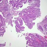
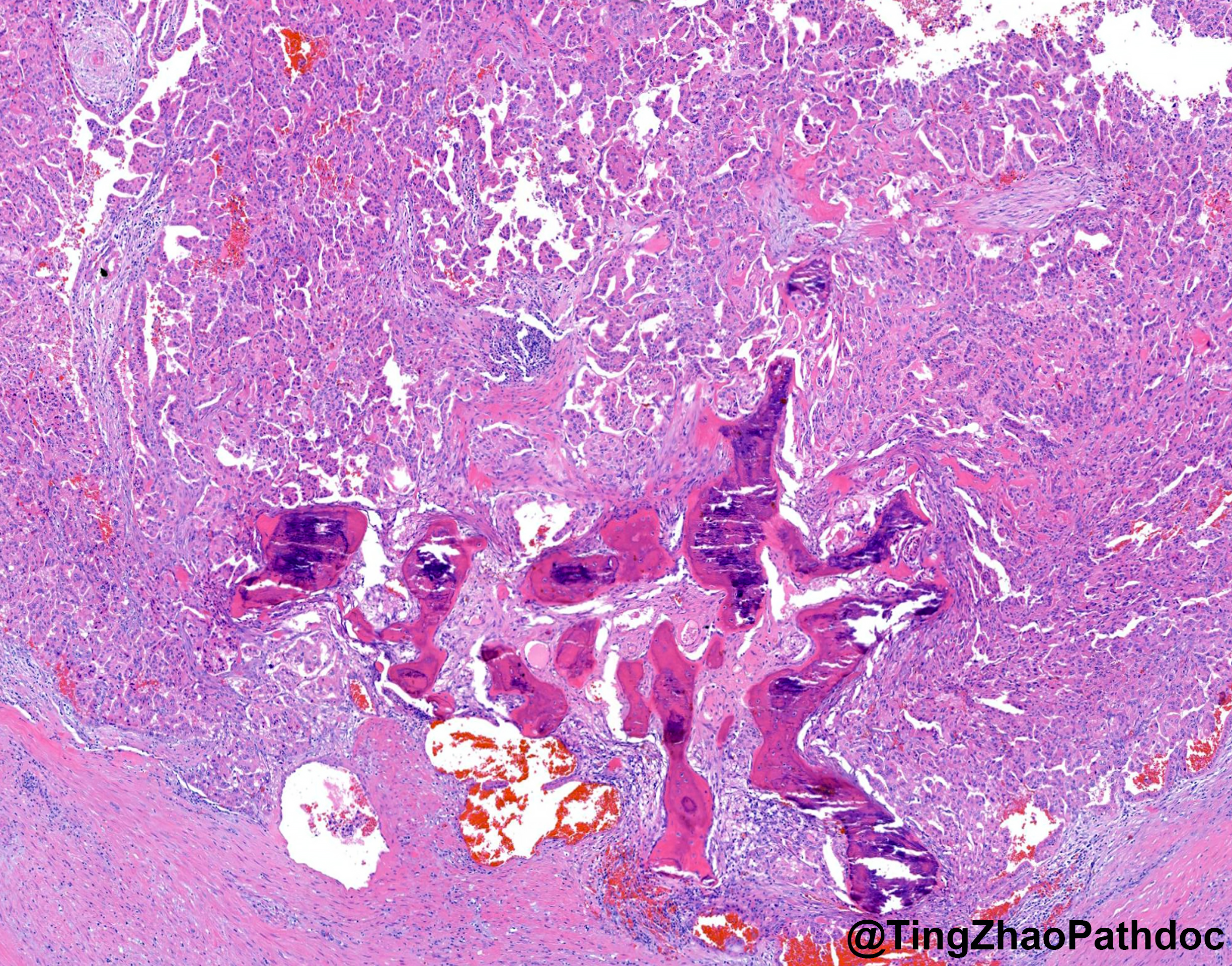
Osseous 🦴 metaplasia in gastric hyperplastic polyp. Super rare !
#GIpath #Pathology #PathX #PathTwitter


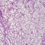
🔬 Clear cell papillary renal cell tumor (CCPRCT):
🎹 Nuclear Polarity
#GUpath #PathX #PathTwitter #Pathology


Ahmed Montasser retweeted

Pathology may be next

Radiology used to have something that was almost invisible because it was so routine:
The reading room consult.
Rounding teams came down.
Surgeons pulled up their cases.
Internists brought the scan that didn't quite fit the clinical picture.
Sometimes the radiologist solved the problem.
Sometimes the clinician supplied one piece of history that completely changed how the images were interpreted.
And usually everyone simply walked away a little sharper.
But something else was happening during all of those conversations.
We were learning each other.
As a radiologist, I learned what individual physicians cared about.
I knew which findings mattered to a particular surgeon.
Which measurements an oncologist was following.
Which details a specialist wanted emphasized.
What questions they were really asking, even when the order just said "pain."
And they learned me.
They knew who was reading their study.
They knew they could walk in, point at something, challenge an interpretation, or ask, "What do you think?"
That relationship created context.
It created feedback.
It created trust.
And over years, it created a kind of institutional knowledge that never appears in the medical record.
When radiology becomes a report produced by someone hundreds or thousands of miles away, we don't just lose proximity.
We risk losing that entire feedback loop.
The report may still be technically excellent.
But the radiologist knows less about the physician.
The physician knows less about the radiologist.
The radiologist sees less of what happened after the report.
And both sides lose opportunities to make each other better.
The reading room was never just a room.
It was where imaging became part of the clinical conversation.
We should think very carefully before designing that conversation out of medicine.
Inspired by a conversation yesterday with @PaleoOnc . Also @909One and @brianchiong .
