

Medicina Crítica Pediátrica
Joined February 2021
- Tweets4.3K
- Following583
- Followers213
- Likes36.3K







Matthew Barriers retweeted

'Look at an arterial pressure trace during mechanical ventilation and you’ll often see the pulse pressure change with each breath. We often refer to it as a ‘swing’. Clinicians recognised this long before monitors began displaying pulse pressure variation (PPV) or stroke volume variation (SVV).
Unlike several indices we’ve examined in this series, PPV and SVV have a sound physiological basis and a substantial body of supporting evidence. Under appropriate conditions, they can predict whether stroke volume will increase following volume expansion.
But there are numerous confounders, and surprisingly few critically ill patients meet all the conditions required for reliable interpretation. Even when the measurements are valid, the physiological behaviour they reveal is not specific to fluid administration. Vasopressors can also increase stroke volume while reducing respiratory variation, without adding blood to the circulation.
What exactly are we measuring, what causes the variation, and what can we conclude from it?'
Read on for free on Substack. Link in reply.


Matthew Barriers retweeted

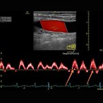
Here’s a continuous-wave Doppler tracing across the tricuspid valve from the right ventricular inflow view.
What’s causing all those horizontal lines?
(The clue is in the inset)
#POCUS #echofirst #Nephpearls

Matthew Barriers retweeted

"Capillary leak" is still a clinical guess. We cannot measure permeability at the bedside, so a multimodal look at edema, congestion, and endothelial injury is the best we have. 🎩 tip to the authors, including @fabio_taccone and @PhilGuerci. eddyjoemd.com/foamed


Matthew Barriers retweeted

Ventilating the injured brain: Balancing lung protection and cerebral physiology
CCR Journal Watch
criticalcarereviews.com/late…


Matthew Barriers retweeted
Setting the vent by tidal volume and plateau alone misses a lot: how hard the patient is breathing, regional lung stress, and the RV. A good review on putting it all together, with us still in charge (for now). 🎩 tip to the authors.
eddyjoemd.com/foamed


Matthew Barriers retweeted

✨ El precio emocional de estar con cada signo del zodiaco ✨
El precio emocional de estar con un Escorpio es que todo lo que dices queda registrado para siempre.
horoscoponegro.com/el-precio…

Matthew Barriers retweeted

The Decisive Test Between Life and Death
In the intensive care unit, the caloric test is performed.. Cold water is injected into the ear, followed by seconds of waiting that feel like ages.
If eye movements are observed: The brainstem is still transmitting signals, and hope remains.
If no response is noted: Brainstem function has ceased, and the last light in the command center has gone out.
Matthew Barriers retweeted

👩🏻⚕️☕️Hoy vamos hablar de:
Understanding the Nuanced Concept of Hemodynamic Incoherence and Its Underlying Physiology
Philippe Rola, Eduardo Kattan, Glenn Hernández et al. (J Clin Med, 2026)
☝🏻🤓Comprendiendo el concepto de incoherencia hemodinámica y su fisiología subyacente🫀
☝🏻🤓Este trabajo redefine el concepto clásico de incoherencia hemodinámica, proponiendo que no se trata de un único fenómeno fisiopatológico, sino de dos entidades con mecanismos completamente diferentes.
👩🏻⚕️☕️Este cambio conceptual tiene implicaciones trascendentales para la reanimación moderna, ya que explica por qué muchos pacientes continúan con hipoperfusión tisular a pesar de alcanzar metas tradicionales de PAM, GC o saturación venosa central.
🔴La perfusión tisular no depende únicamente del flujo sanguíneo global.🫁🧠🫀
🔴Depende de la capacidad del sistema cardiovascular para transferir energía hidráulica desde el ventrículo izquierdo hasta la mitocondria atravesando múltiples interfaces vasculares.‼️
☝🏻🤓Cuando alguna de estas interfaces falla, aparece la incoherencia hemodinámica, donde:
🔸️PAM normal, GC adecuado
, Vasopresores suficientes
⚠️NO significan necesariamente adecuada oxigenación celular.
☝🏻🤓El nuevo modelo fisiológico: las cuatro interfaces hemodinámicas🫀
Rola integra el concepto desarrollado previamente por Hernández y Kattan de las 4 interfaces de la circulación:
🔴Interface I Ventrículo izquierdo → árbol arterial
(Acoplamiento ventrículo-arterial)
👇🏽
🔴Interface II Arteriolas → capilares
(Control de la resistencia precapilar)
👇🏽
🔴Interface III Capilares → sistema venoso.
(Drenaje venoso y presión venosa)
👇🏽
🔴Interface IV Ventrículo derecho → circulación pulmonar
(Acoplamiento VD-arteria pulmonar)
☝🏻🤓Cada una puede convertirse en un punto de pérdida de energía y generar hipoperfusión aun cuando el resto del sistema funcione correctamente.
🫀Tipo 1️⃣: Incoherencia microcirculatoria primaria
🔹️Es la forma clásica descrita por Ince.
🧐Identifiquemos el problema👀 Aquí el problema está dentro de la microcirculación.‼️
Y ...Se caracteriza por: 🔹️Heterogeneidad capilar.
🔹️Shunts funcionales.
🔹️Glucocálix lesionado.
🔹️Alteración endotelial.
🔴Hay una pérdida importante de la autorregulación👀‼️
🚿Edema intersticial.
☝🏻🤓El mejor ejemplo es la sepsis.
➡️Aunque el gasto cardíaco sea elevado y la PAM sea adecuada, el flujo capilar continúa siendo extremadamente heterogéneo.
🧐Resultado:
→ Persistencia de hipoxia celular.
🫀Tipo 2️⃣: Incoherencia secundaria
🧐Aquí la microcirculación es potencialmente normal, pero no recibe suficiente energía hidráulica debido a un problema "aguas arriba" o "aguas abajo".
Y...Puede producirse por:
🔹️Bajo gasto cardíaco.
🔹️Vasoconstricción excesiva.
🔹️Hipertensión venosa
🔹️Congestión
🔹️Disfunción del ventrículo derecho
🔹️Desacoplamiento ventrículo-arterial🫀
⚠️En este escenario el problema no es el capilar, sino la transmisión del flujo.
💎Esta clasificación cambia completamente la estrategia terapéutica.
🫀Relación con la perfusión tisular
🎯El objetivo de la reanimación deja de ser:🚫 "subir la presión"...y pasa a ser: "Restaurar la transferencia de energía hacia la célula."🆗️
🔴Por ello adquieren importancia marcadores como:
☝🏻Tiempo de llenado capilar (CRT)
🔹️Moteado cutáneo
➡️Lactato
🔴Diferencia venoarterial de CO₂.
🔹️Relación P(v-a)CO₂/C(a-v)O₂
🚿VExUS
🫀Ecografía Doppler
👅Microcirculación sublingual (cuando esté disponible).
💎💎Relación con el acoplamiento ventrículo-arterial
🤓☝🏻Uno de los conceptos más interesantes del artículo es que el acoplamiento ventrículo-arterial (VA) constituye la primera interfaz.
🫀Cuando existe desacoplamiento:
🔹️Disminuye la eficiencia energética
🔹️Cae el volumen sistólico efectivo
🔹️Aumenta el costo miocárdico
🔹️Disminuye la energía transmitida a la microcirculación.
@AndromedaShock
@edu_kattan
👇🏽👇🏽 – at Benito Juárez, Distrito Federal


Matthew Barriers retweeted
Today's Paper of the Day is:
Unplanned postoperative reintubation: a narrative review of risk factors, predictive tools, and preventive strategie
criticalcarereviews.com/late…
Join us to read 1 paper per day and stay up-to-date as we cover the spectrum of critical care across 2026

Matthew Barriers retweeted
Triple M overlap syndrome in the intensive care unit: differential diagnosis and management
CCR Journal Watch
criticalcarereviews.com/late…


Matthew Barriers retweeted

Pulmonary hypertension 🫁 + LEFT heart disease🫀: common, complex, and STILL full of unanswered questions. Read about emerging strategies in the latest #JACC review: jacc.org/doi/10.1016/j.jacc.…
#PulmonaryHypertension @GreggWStone @yreddyhf @mardigomberg @ShahzebKhanMD

Matthew Barriers retweeted

Critical Physiology Series #16
LVEDV, E/e′ and PCWP: Volume Is Not Pressure
When evaluating preload in critically ill patients, one of the most common mistakes is assuming that ventricular volume and filling pressure are the same. They are not.
LV end diastolic volume (LVEDV) tells us how much blood is inside the ventricle before systole. E/e′ and pulmonary capillary wedge pressure (PCWP) estimate the pressure required to fill that ventricle.
A healthy, compliant ventricle can accommodate a large volume with little increase in pressure. A stiff ventricle, such as in diastolic dysfunction or sepsis related myocardial dysfunction, may develop very high filling pressures despite having a normal or even small LVEDV.
This distinction is crucial in the ICU.
A patient with a small LV cavity does not always need fluids. If E/e′ is elevated, the ventricle may already be under high filling pressure, and additional fluids may increase pulmonary congestion without improving cardiac output.
Likewise, an elevated PCWP reflects increased left sided filling pressure, not necessarily adequate preload or fluid responsiveness.
The bedside message is simple.
LVEDV estimates volume.
E/e′ and PCWP estimate pressure.
Neither predicts fluid responsiveness alone. They should always be interpreted together with dynamic tests and the overall clinical picture.
Recommended lectures 📚
Bouhemad, 2003, doi.org/10.1097/00000542-200…
Nagueh, 2016, doi.org/10.1093/ehjci/jew082

Matthew Barriers retweeted
My mental model for approaching #AKI - the framework I encourage residents to use when presenting cases on their #nephrology rotation.
Once you interpret the urinalysis and #POCUS findings [see thread] (or other imaging if available), you’ve already done most of the diagnostic work.
