
Alone we go faster, together we go further
Beaujon Hosp, Clichy, France
Joined May 2021
- Tweets23
- Following98
- Followers51
- Likes52

Beaujon Radiology retweeted

CT to raise suspicion of Porto-Sinusoidal Vascular Disorder (PSVD)
✅Liver Surface Nodularity < 2.5
➕
✅ No atrophy of segment 4
👉 > 90% specificity for PSVD
@HEP_Journal @RautouE @jcgarciapagan
aasldpubs.onlinelibrary.wile…

Beaujon Radiology retweeted
Watching the never ending list of "impact of Covid-19 on ..." papers I'm surprised we don't already have the "impact of Covid-19 on the management of patients with Covid-19" study...
Beaujon Radiology retweeted
HUGE update of the @BCLC_group in @JHepatology @ma5RN @Alexfornergon @bruixj @JordiRimola @docamitgs @riadsalemIR
✅Better pts characterization (BCLC-B ++)
✅Help for clinical decision making
✅ Radioembolization does exist!
✅ BCLC progression algo
tinyurl.com/2ahmxsvu

Beaujon Radiology retweeted
TACE for liver NET metastases (222 patients)
✅Median TTLP 19.3 mos
✅Median OS 5.3 yrs
✅OS ⬆️ in mRECIST responders (80.5 vs. 39.6 mos).
✅ Age, grade, liver involvement predict worse OS
Awesome study in @MDPIOpenAccess @GustaveRoussy @Beaujon_Rad mdpi.com/2072-6694/13/21/536…

Beaujon Radiology retweeted
Cystic Fibrosis-related liver disease (2/2)
“red-flag” features to suspect PSVD in pts with PHT
1⃣ no liver anomalies
2⃣ LSM < threshold for cirrhosis
3⃣ coarse macronodular pattern
Great review by J. Dana, V. Vilgrain @Beaujon_Rad in @JHepatology tinyurl.com/3y3vk6ck
Beaujon Radiology retweeted
Cystic Fibrosis-related liver disease (1/2)
✅ PHT=main complication due to cirrhosis or PSVD
✅ US=key for diagnosis + follow-up
✅ Liver stiffness measurement may be helpful
Great review by J. Dana, V. Vilgrain @Beaujon_Rad in @JHepatology tinyurl.com/3y3vk6ck #livertwitter
Beaujon Radiology retweeted
Colonic involvement in Acute Mesenteric Ischemia
1⃣ found in 28%
2⃣ most common in the right colon
3⃣ increased morbidity (SBS, PS) and mortality
➡️ should be searched for on CT!
@inteststroke @AlexandreNuzzo @oliviercorcos @Beaujon_Rad @dr_manuweiss
link.springer.com/article/10…
Beaujon Radiology retweeted
Follow up of benign nodules in Budd Chiari or after Fontan with MRI
✅ 40% disappear
✅ 45% change in size and/or signal intensity
✅ changes in BCS >> Fontan
➡️ dynamics to be aware of !
@marco_dioguardi @RautouE @Beaujon_Rad #livertwitter
sciencedirect.com/science/ar…

Beaujon Radiology retweeted
In patients with #NOMI no bowel enhancement, bowel thinning, bicarbonate ≤15 mM and PT <40%
1⃣Stratify the risk of bowel necrosis
2⃣> @inteststroke score by @AlexandreNuzzo
Great study by P. Calame in @AmJGastro
Proud to be part of it !
pubmed.ncbi.nlm.nih.gov/3418…

A walk along the path of imaging for the diagnosis and prognosis of #HCC with @Riccard49700689 @Jules_GREGORY_ @marco_dioguardi and @maximeronot published in @LiverInt Stimulating !! onlinelibrary.wiley.com/doi/…
55-yo male patient with chronic HBV infection but no cirrhosis. Surveillance US exam shows a 20mm nodule in the left liver lobe. The patient undergoes MRI for characterization. Simple question : am I allowed to call or an HCC ? @RadiologieUNIR #liver

Check the super useful presentation by @Marco_dioguardi about tips and tricks to access the inaccessible lesions at #Esgar2021. Available on-demand
Today @VChernyakMD and @ClaudeSirlin (with a bit of help from #Shakespeare) gave the most impressive, fun and educational duet lecture about #LIRADS at #esgar2021 Make sure to watch it’ available on-demand
Make sure to watch the great post-graduate course on Benign liver tumors with Valérie Vilgrain @giubrancatelli @valerie_paradis and Raneem Albazaz at #esgar2021. Available on-demand !!
Beaujon Radiology retweeted

#surveillance #HCC #MRI Good article about "Alternative approach of hepatocellular carcinoma surveillance: abbreviated MRI" from Dr. Robert M. Marks's team. @RyanBrunsing @chemshift1 @bobmarks76 @ClaudeSirlin @VChernyakMD
Download for free: hrjournal.net/article/downlo…

Teduglutide induces a significant SB wall thickness ⬆️ that can be depicted by imaging <6 mo after treatment initiation. The degree of such ⬆️ may be associated with clinical response. Check out study in @AJCNutrition with @maximeronot and @franciscajoly academic.oup.com/ajcn/articl…
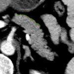
Found of quantitative imaging? We are too! With @Riccard49700689 we quantified the Pancreas Surface Lobularity on CT and found it to ⬆️ with age and BMI and to be > in men. Stimulating perspectives. Check out study in KJR kjronline.org/DOIx.php?id=10…


Serotonin+ pancreatic #NET ⬆️fibrous stroma, small but marked MPD dilation and ⬆️ RFS after surgery. Check our new study in European Radiology by Marco Dioguardi Burgio with @louis_demestier @maximeronot link.springer.com/article/10…