

Flagship journal of @Intlsocdiff. Published by #Elsevier for 40+ years. All things relating to differentiation, development, stem cells and regeneration
- Tweets248
- Following331
- Followers350
- Likes144
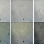



ALT Fig. 2. A. MSC on day 3. Cultures were terminated on day 3 as Mesenchymal Stem Cells (MSCs) achieved confluency, a point beyond which further culture was not feasible. B. Res-only group on day 21. C. 5-aza group on day 21. D. Res 5 group on day 21. E. Res 10 group on day 21. F. Res 20 group on day 21. Perinuclear clear regions that appeared to be consistent with cytoplasmic lipid droplet accumulation are indicated by black arrows, and cell-to-cell junctions morphologically resembling intercalated disc-like structures are highlighted by a black circle. The magnification ×20.

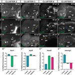
ALT Fig. 3. Target genes from each cluster are differentially expressed in prdm3 and prdm16 mutant zebrafish.

ALT Fig. 2. Prof. Sally Dunwoodie presented awards to the winners of the best poster presentation.

ALT xZnf131-depleted embryos exhibit gastrulation defects

ALT Fig. 2. Model of endocardial cushion EndMT. This figure illustrates the developmental progression of endocardial cushion EndMT. Firstly, the cardiac jelly is generated between the endocardium and myocardium. Secondly, endocardial cells (ECs) sense blood flow shear forces, lose their primary cilia, and initiate EndMT. ECs detach from the endocardium, migrate into the cardiac jelly, and transform into mesenchymal cells. During post-EndMT development, mesenchymal cells undergo proliferation, migration, and secretion of extracellular matrix, participating in the shaping of valves and the atrioventricular septum.

ALT Fig. 1. Clinical and radiological features of the proband with acromesomelic dysplasia, Maroteaux type (AMDM).

ALT Fig. 3. Image Analysis reveals proliferation differences based on differentiation stimuli. (A) Confluency seen within each well is graphed over the measurement period. (B) Representative images comparing macrophages at day 0 and day 18, with the differentiation agent concentration increasing down the image pairs.


ALT Fig. 4. Table of all sgRNAs designed and tested in this study

ALT Fig. 7. Proposed model for promoter-level reciprocal repression during Xenopus gastrulation. In ventral mesodermal regions, BMP signaling activates Ventx1.1 expression through Smad1/5/8–Smad4 signaling. Ventx1.1 represses gsc transcription through VRE elements within the proximal gsc promoter. In the neuroectodermal domain, reduced BMP signaling permits Foxd4l1.1 expression, which further represses gsc transcription through BRE/FRE-containing promoter region. In dorsal organizer regions, Activin/Nodal signaling activates gsc through Smad2/3–Smad4 signaling, and Gsc represses ventx1.1 transcription through the GRE1-containing region of the ventx1.1 promoter. The FRE partially overlaps with VRE2 on the reverse strand, suggesting spatially distinct promoter occupancy by Ventx1.1 and Foxd4l1.1 during gastrulation.

ALT Fig. 1. Sagittal histological section of the developing foot showing the anatomical localization of the human heel fat pad (HHFP) (CRL 177 mm; 19 GW). The calcaneus and overlying skin are identified. The inset highlights the region of interest for HHFP development. Scale bar: 100 μm.

ALT Fig. 1. DEL1 is highly expressed and secreted during osteoclast differentiation.

ALT Fig. 6. Proposed model of the IHH signaling pathway modulation by RAB23-GDP, -GTP and -Resveratrol molecular interactions. The rendered images represent the functional state of RAB23 protein (active or inactive) according of molecular docking with GDP, GTP and Resveratrol, and its impact in the IHH gene transcription (promotion or inhibition) induced by GLI1 transcription factor, in l-PDLCs cultivated in osteogenic medium with and without resveratrol pretreatment (OM and OM/RESV groups, respectively).


ALT Call for papers! Submit to the Splicing in Development and Disease special issue by June 30, 2026.