- Tweets42
- Following27
- Followers85
- Likes50


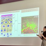




ALT Confocal fluorescent images of tachyzoites expanded by U-ExM while undergoing helical gliding by crossing over anti-adhesive 4 µm wide PLL-PEG lanes that separate fibronectin-Atto488-coated rectangles (2 µm, yellow areas). Each frame recapitulates shows tachyzoites immunostained for the submembranous GAP45 glideosome component (green) and for the cortical microtubules (magenta).


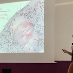
ALT The Toxoplasma parasite (green contour and blue nucleus) produces a rosette of progeny inside a human cell (image center). The progeny then egresses in a helical gliding sequence ( image periphery) during which the parasite successively bends and extends supported by the compressed or straight microtubule cytoskeleton (green) that emerges at the apical pole. The myosin motor (pink) contributes to the traction force during apical bending. U-ExM sample.