
Stanford - Radiology AI | RadAI @ HOPPR | Previous: ML @HuggingFace, Academic staff - Research @Stanford University, @StanfordAIMI Affiliate
Palo Alto
Joined July 2017
- Tweets306
- Following939
- Followers1.2K
- Likes219





#RadiologyAI Our report drafting solution is deployed in 300 sites over 3 countries.
Here are 𝘁𝗵𝗲 𝗼𝗽𝗲𝗻 𝗾𝘂𝗲𝘀𝘁𝗶𝗼𝗻𝘀 we faced when building VLMs that scale across healthcare systems, radiologists of different nationalities, reporting styles, clinical use cases, and varying finding prevalence.
This effort created 𝘂𝗻𝗽𝗿𝗲𝗰𝗲𝗱𝗲𝗻𝘁𝗲𝗱 𝗠𝗟 𝗰𝗵𝗮𝗹𝗹𝗲𝗻𝗴𝗲𝘀: how can a single foundation model satisfy radiologists when one reports aortic knob prominence 90% of the time, while their colleague next door reports it 0% of the time?
After all, we have all adapted to using the same ChatGPT or Claude. Radiologists, however, are not ready to adapt to a single, standardized threshold for reporting findings. 𝗧𝗵𝗲 𝗺𝗼𝗱𝗲𝗹 𝗺𝘂𝘀𝘁 𝗮𝗱𝗮𝗽𝘁 𝘁𝗼 𝘁𝗵𝗲𝗺, 𝗻𝗼𝘁 𝘁𝗵𝗲 𝗼𝘁𝗵𝗲𝗿 𝘄𝗮𝘆 𝗮𝗿𝗼𝘂𝗻𝗱.
𝗔𝗻𝗱 𝘁𝗵𝗮𝘁 𝗶𝘀 𝗽𝗲𝗿𝗳𝗲𝗰𝘁𝗹𝘆 𝗳𝗶𝗻𝗲. But to do so, you need to understand many overlooked element in the NLP/NLG scientific literature:
- How do you control a model's conservatism or verbosity through metadata?
- How do you define an operating point, a decision boundary in language space, that determines whether a finding should be reported, especially when generating an entire radiology report section?
- How do you train a decoder to predict whether a radiologist will sign off on a generated report?
- And most importantly, how do you truly understand your end user: the radiologist?
At Eden, our technology stack is already deployed across 𝗺𝗼𝗿𝗲 𝘁𝗵𝗮𝗻 𝟮,𝟴𝟬𝟬 𝗶𝗺𝗮𝗴𝗶𝗻𝗴 𝗱𝗲𝗽𝗮𝗿𝘁𝗺𝗲𝗻𝘁𝘀 𝗶𝗻 𝟮𝟬 𝗰𝗼𝘂𝗻𝘁𝗿𝗶𝗲𝘀, giving us a unique opportunity to solve these challenges at scale.
#RadiologyAI New proceedings alert 🚨 . Finally, someone does the painstaking groundwork. 𝗡𝗜𝗛’𝘀 𝗖𝗧-𝗕𝗲𝗻𝗰𝗵 𝗽𝘂𝘁𝘀 𝘄𝗼𝗿𝗱𝘀 𝗼𝗻 𝟮𝟬,𝟯𝟯𝟱 𝗖𝗧 𝗹𝗲𝘀𝗶𝗼𝗻𝘀 (𝗠𝗘𝗟𝗕𝗔 𝟮𝟬𝟮𝟲). 𝗢𝗻 𝗶𝘁𝘀 𝗳𝗼𝘂𝗿-𝗰𝗵𝗼𝗶𝗰𝗲 𝗲𝘅𝗮𝗺, 𝗚𝗣𝗧-𝟰𝗩 𝘀𝗰𝗼𝗿𝗲𝘀 𝟮𝟴%.
CT-Bench is an exam for AI on single CT lesions: which of these four descriptions fits this lesion? Which image matches this description? How big is it?
Picking at random scores 25%. GPT-4V scores 𝟮𝟴%. It is a 2023–2024 model; nothing newer was tested.
𝗔𝗻𝗱 𝗿𝗮𝗱𝗶𝗼𝗹𝗼𝗴𝗶𝘀𝘁𝘀? 𝗧𝘄𝗼 𝘀𝗲𝗻𝗶𝗼𝗿 𝗿𝗮𝗱𝗶𝗼𝗹𝗼𝗴𝗶𝘀𝘁𝘀 𝗴𝗼𝘁 𝗮𝗯𝗼𝘂𝘁 𝟴 𝗶𝗻 𝟭𝟬 𝗿𝗶𝗴𝗵𝘁, 𝗼𝗻 𝟭𝟬𝟬 𝗰𝗮𝘀𝗲𝘀.
→ The best AI in the paper, a model trained on the released lesions, gets 𝟱𝟬% overall.
→ Draw a box around the lesion and that model goes from 37% to 62%. The senior radiologists go from about 7 in 10 to about 9 in 10. Knowing where to look matters for both.
𝗪𝗵𝗮𝘁 𝘆𝗼𝘂 𝗰𝗮𝗻 𝘂𝘀𝗲 𝘁𝗼𝗱𝗮𝘆:
→ 𝟮𝟬,𝟯𝟯𝟱 𝗖𝗧 𝗹𝗲𝘀𝗶𝗼𝗻𝘀, each with a one-sentence description, a size and a box, plus 2,850 exam questions with the paper’s scores to compare against. On Kaggle (link in the first comment).
→ The licence is CC BY-NC-SA 4.0: free for research, 𝗻𝗼 𝗰𝗼𝗺𝗺𝗲𝗿𝗰𝗶𝗮𝗹 𝘂𝘀𝗲.
The lesson: before trusting a model with a whole report, test it lesion by lesion: 𝘄𝗵𝗲𝗿𝗲 𝗶𝘁 𝗶𝘀, 𝘄𝗵𝗮𝘁 𝗶𝘁 𝗶𝘀, 𝗵𝗼𝘄 𝗯𝗶𝗴 𝗶𝘁 𝗶𝘀.
Public CT data that ties a sentence to a single lesion is rare, and this set is a download away.
Full breakdown in the first comment 👇
Blog post: jbdel.github.io/research-blo…
Paper: melba-journal.org/papers/202…
Data: kaggle.com/datasets/cd1661d6…

#RadiologyAI My highlight of #MICCAI2026 𝗟𝘂𝗻𝗶𝘁’𝘀 𝗦𝗣𝗘𝗖-𝗖𝗫𝗥 𝗴𝗿𝗮𝗱𝗲𝘀 𝗔𝗜 𝗫-𝗿𝗮𝘆 𝗿𝗲𝗽𝗼𝗿𝘁𝘀 𝗳𝗶𝗻𝗱𝗶𝗻𝗴 𝗯𝘆 𝗳𝗶𝗻𝗱𝗶𝗻𝗴. 𝗧𝗵𝗲 𝗰𝗮𝘁𝗰𝗵: 𝗼𝗻 𝗺𝗼𝘀𝘁 𝗸𝗶𝗻𝗱𝘀 𝗼𝗳 𝗳𝗶𝗻𝗱𝗶𝗻𝗴, 𝘁𝗵𝗲 𝗯𝗲𝘀𝘁 𝗔𝗜 𝗶𝘀 𝗺𝗼𝘀𝘁𝗹𝘆 𝘄𝗿𝗼𝗻𝗴.
SPEC-CXR asks four questions about every finding: is it there, in the right place, with the right severity, and compared correctly with the last X-ray?
Tubes and devices get found. But on 𝟵 𝗼𝗳 𝟭𝟱 kinds of finding, the best model, Med-Gemini, either misses at least 4 in 5 real cases or gets at least 4 in 5 of its calls wrong.
𝗪𝗵𝗮𝘁 𝘆𝗼𝘂 𝗰𝗮𝗻 𝘂𝘀𝗲 𝘁𝗼𝗱𝗮𝘆:
→ The code (link in the first comment), free for non-commercial research.
→ The checklist of 122 findings and diagnoses, as plain files you can reuse (same licence) for your own per-finding table.
→ One rule: name your AI judge and keep it fixed. Across the paper’s eight judges, agreement with radiologists ranges from 𝟬.𝟲𝟬 𝘁𝗼 𝟬.𝟳𝟴.
Caveat: the finding-level grades were checked on only 50 report pairs, and the released code runs a different version of the pipeline from the one the paper validated.
The lesson: 𝗼𝗻𝗲 𝘀𝗰𝗼𝗿𝗲 𝗽𝗲𝗿 𝗿𝗲𝗽𝗼𝗿𝘁 𝗰𝗮𝗻’𝘁 𝘀𝗮𝘆 𝘄𝗵𝗶𝗰𝗵 𝗳𝗶𝗻𝗱𝗶𝗻𝗴𝘀 𝗮 𝗺𝗼𝗱𝗲𝗹 𝗺𝗶𝘀𝘀𝗲𝘀. Grade findings, not just reports.
Still, SPEC-CXR is a gift to the field: a ready checklist and released code to do exactly that.
Disclosure: I’m a co-author of GREEN and RadGraph-F1, two of the metrics this paper positions itself against.
Full breakdown in the first comment 👇
Blog post: jbdel.github.io/research-blo…
Paper: papers.miccai.org/miccai-202…
Code: github.com/lunit-io/spec-cxr

1
56
#MICCAI2025 HIGHLIGHTS #RadiologyAI 𝗖𝗵𝗲𝗰𝗸𝗶𝗻𝗴 𝗮𝗻 𝗫-𝗿𝗮𝘆 𝗿𝗲𝗽𝗼𝗿𝘁 𝗮𝗴𝗮𝗶𝗻𝘀𝘁 𝘁𝗵𝗲 𝗶𝗺𝗮𝗴𝗲, 𝗻𝗼𝘁 𝗮 𝗿𝗲𝗳𝗲𝗿𝗲𝗻𝗰𝗲. A paper reports 0.997 agreement with ground truth.

• No code or model listed; omitted findings are out of scope.
Great work by Razi Mahmood and co-authors (RPI, IBM Research, Stanford, MGH)
1
14
Blog post: jbdel.github.io/research-blo…
Paper: papers.miccai.org/miccai-202…
Data: huggingface.co/datasets/razi…

17
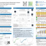
#RadiologyAI at #NeurIPS2025
Interested in generating radiology reports? This work does the grunt, un-sexy work: modeling uncertainty.
The key insight is simple but powerful:
trustworthy AI doesn’t come from bigger models; it comes from explicitly modeling uncertainty and doing the hard reasoning work.
CURV's idea is to flip that:
explicitly label and ground uncertainty: what’s unclear, where it is, and why.


1
265
#RadiologyAI at #neurips2025
OK, I love this one.
This is not, at first, radiology-related. But this one trick could absolutely translate to medical imaging. For architectures that don't use full self-attention, 𝗵𝗼𝘄 𝘆𝗼𝘂 𝗼𝗿𝗱𝗲𝗿 𝗽𝗮𝘁𝗰𝗵𝗲𝘀 𝗶𝗻 𝘆𝗼𝘂𝗿 𝗩𝗶𝗧 𝗮𝗰𝘁𝘂𝗮𝗹𝗹𝘆 𝗺𝗮𝘁𝘁𝗲𝗿𝘀.
Think about a chest x-ray, why should patches be read left-to-right, top-to-bottom like a PDF? Anatomy isn't linear. For chest X-rays, clinically meaningful context follows organs, symmetry, and spatial relations. If your ViT uses local or sparse attention, patch ordering becomes an inductive bias and we should design it to match anatomy.
Patch order ≠ implementation detail.
If you want to try this on open-source x-ray datasets, pm me 😉

2
211
#RadiologyAI at #neurips2025
So nice to see dinotxt has been applied to chest x-rays. Its actually an idea I wanted to try for a long time :-). Zero-shot segmentation is seriously impressive, and this paper shows you can precisely ground findings directly in the chest x-ray. Pretty neat...


5
249