

Senior Medical Scientist, Electron Microscopy Unit, SA Pathology, Adelaide, Australia. Ultrastructural pathology of kidney, muscle, nerve, liver, tumours, etc.
Australia
Joined April 2019
- Tweets898
- Following1.4K
- Followers1.4K
- Likes2.2K

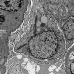


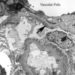
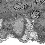
Renal tx insertion bx. @electronmicroscopy shows a rare finding of ribosome-lamella complexes (arrows) in podocytes. Does anyone know if they are significant? I think it's a non-specific change reflecting cell stress. Not to be confused with crystals that can occur in podocytes.


Renal biopsy of a patient with T1DM. #electronmicroscopy of medullary tubules. Severe accumulation of glycogen in the epithelium. The nuclei (easily misinterpreted as vacuoles) contain so much glycogen that the chromatin and nucleoli have been pushed to the nuclear periphery.


These structures are seen by @electronmicroscopy in smooth muscle cells of kidney arteriolar walls and juxtaglomerular cells. These 'lipofuscin-like granules' should not be misinterpreted as inclusions of the type found in some forms of neuronal ceroid lipofuscinosis.


#askrenal Can a plasmacytoma produce anti-LCAT autoantibodies?
We have a case of a patient with a plasmacytoma. However, by #electronmicroscopy, the glomerular ultrastructure suggests LCAT deficiency.
#electronmicroscopy Renal tx bx from a child w renal dysplasia as native disease. Any thoughts on these structures in tubules - ? lipofuscin, ? peroxisomes, ? a drug reaction. It’s the structures w periodicity that interest me. Human peroxisomes typically lack a crystalloid core


#electronmicroscopy Kidney bx shows a membranous nephropathy. #podocytes, typically post-mitotic, appear to be trying to divide. Nuclei are bi-lobed with narrow chromatin bridges. Cells are rounded with blebs + detachment from the GBM. Possibly an example of mitotic catastrophe.


Native Kidney Bx #electronmicroscopy. #askrenal Any thoughts on these electron-dense structures? I interpret them as karyorrhectic debris. I see them rarely but have never seen them documented in an illustration. Interestingly this patient has 1+ C1q in capillary walls.


Kidney Bx. Looking for an infective agent by #electronmicroscopy.
Found these structures that can be misinterpreted as viruses. They are nuclear pores. The plane of section has tangentially traversed the edge of a nucleus.


Patient w membranous nephropathy. #electronmicroscopy shows electron-dense deposits (arrow) lining Bowman's capsule (BC) at the vascular pole, but deposits are not evident further along BC. Presumably the transition point between #podocyte and parietal epithelial cell phenotypes.


Native kidney bx of a 50M w psoriasis. @electronmicroscopy shows membranous nephropathy (MN). Is there a relationship between psoriasis and MN? Could there be a PLA2 receptor auto-antibody that attacks both keratinocytes in skin + podocytes in glomeruli? @SethiRenalPath #askrenal


Last chance to register for the Electron Microscopy Tutorial Day & UltraPath XXI Conference, Sep 30-Oct 4. You can't beat the location. Follow the pink arrow and you'll find us discussing ultrastructural pathology at Circular Quay in the heart of Sydney.
ultrapath.org

Are Langerhans histiocytes a normal component of the renal tubular epithelium? We have patient w uveitis + minimal inflammatory infiltrate on renal bx. EM shows structures that look like Birbeck granules (arrows). Could the presence of Langerhans cells indicate anything?#askrenal


Renal Bx, female in 40s. TBMs focally expanded by fibrils measuring 12-13nm in diameter. I see this occasionally by EM in renal biopsies and never know if it's amyloid or a mimic (similar to diabetic fibrillosis in mesangial matrix). No amyloid in gloms. Congo Red not done ATM.


Seeking opinions for a differential on this appearance by #electronmicroscopy. Native Renal Bx, male in 30s.
My thoughts are LCAT Deficiency or Alagille Syndrome. Any others? - ? hepatic glomerulosclerosis. Pt has low HDL. #askrenal @SethiRenalPath @ChangUCanSpare @JZRenalPath


Seeking opinions on this subendothelial material. Native Renal Bx. Male in 60s w hematuria + proteinuria. LM shows an MPGN w focal vasculitis. EM is from the paraffin block - occasional loops occluded. IF - focal staining for fibrinogen. ? cryofibrinogen
#askrenal @SethiRenalPath


Please consider attending the UltraPath XXI electron microscopy conference in beautiful Manly Beach, Sydney, Sep 30 to Oct 4, 2024. Lots of interesting ultrastructural pathology with presentations by world leaders in the field. Plus great social events.
ultrapath.org/


Alveolar Soft Part Sarcoma
Has any progress been made in uncovering the histogenesis of this tumour?
#askrenalpath @AstridWeins
Do patients with anti-nephrin autoantibodies still show slit diaphragms between intact podocyte foot processes by #electronmicroscopy?