

Pioneering end-to-end solutions for 3D spatial biology.
Cambridge, MA
Joined June 2018
- Tweets556
- Following408
- Followers535
- Likes365



We got to talk about the essentials of sample prep for light sheet imaging at @WesternuWIN's Mesoscopic Integrated Neuroimaging Data (MIND) platform, including how to fast-track projects with their SmartBatch+ device. Thank you for hosting us! #LightSheet #TissueClearing


How does the cortex help regulate sleep & wakefulness? Researchers have ID'ed a sparse population of inhibitory neurons with unusually long axons which increase synchronization across multiple neocortical regions & promote sleep: bit.ly/47TH91Y #FluorescenceFriday
1 month left to apply for our #SfN26 travel award! We want to hear how you used the LifeCanvas 3D discovery pipeline in your poster. No form required – just post an image or video representing your research and tag us. We'll share + include your research in our SfN poster guide!
📹 This #FluorescenceFriday video is courtesy of @ShreyasMSurya, showing neurons projecting from the visual cortex to different parts of the brain. Directly generated neurons are shown in red, and indirectly generated neurons are shown in green.
This #FluorescenceFriday video from @KenIshii1223 shows mu-opioid receptor expressing cells (red) & c-FOS signal (green) in a mouse brain after exposure to acute morphine. Learn about the science behind this SmartSPIM light sheet data in our 11/5 webinar: bit.ly/4ri944r
Apps for @MALifeSciences's ARC awards are due 11/6! Thanks to this program, we're working with the Laboratory of Systems Pharmacology @ Harvard Med to develop high-plex 3D imaging methods to advance cancer research. We're always open to new collabs: bit.ly/4rELfUG
Is it really that easy to get 3D data from a 12 well plate? For #MicroscopyMonday, we thought we’d let #DALISPIM co-creator Nick Evans show you how our open-top light sheet microscope works, from plating samples to acquiring high-res data. Learn more: bit.ly/49pqjKd
#SmartSPIM light sheet microscopy is coming to Barcelona! We can't wait to show you how easy it is to capture hi-res 3D data throughout your entire sample, like this intact mouse brain and spinal cord, mouse duodenum, and rat heart. #FluorescenceFriday
Thank you for the featured data:
Mouse brain - @ShreyasMSurya (Huang lab, Duke)
Mouse duodenum - Dr. Hanan Mohammed (Ramadi lab, NYU Abu Dhabi)
Rat heart - Dr. Joshua Harrison (Birren lab, Brandeis)
@SPAOM5 @REMOABioimaging @PPBioImaging
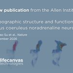
Scientists at the @AllenInstitute have made major strides in understanding one of the brain’s key neuromodulatory systems. Read on to learn more and check out the open access paper... 🧵(1/5)

This work marks a significant advancement in our understanding of the noradrenaline system, with implications for how we treat mood and attention disorders. 📖 Read it here: nature.com/articles/s41586-0… (5/5)
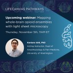
We're kicking off a new webinar series with Dr. Ken Ishii from @gstuber's lab! Join us 11/5 to learn how Dr. Ishii used LifeCanvas tools to create the first cellular-resolution whole-brain atlas of opioid administration and withdrawal states: bit.ly/472YiFV #SmartSPIM

It’s that time of year: #SFN26 abstract acceptances are in, and we want to hear about yours! Entering is easy: just tag us in a post about how you used LifeCanvas tools in your poster. Read on to learn about these clips from last year.. @SfNtweets 🧵(1/3)
0:00-0:19 - Mouse bladder imaged with SmartSPIM by @ZhennWang (Brian S. Kim's lab, Icahn School of Medicine at Mount Sinai). Read Dr. Wang's latest preprint on interorgan neuroimmune circuitry: researchsquare.com/article/r… @itchdoctor (2/3)
0:20-0:35 - Mouse lumbar spinal cord imaged with SmartSPIM by Dr. Andrew Crowther (Virginia Commonwealth University).
0:36-0:48 - Mouse brains at different developmental stages, imaged with SmartSPIM by @ingvildeb (@yongsookimlab, Penn State College of Medicine).
(3/3)