

Pediatrics Challenging Questions...please call everyone interested to follow us.All are cases for discussion and learning .🩺.
Joined October 2024
- Tweets6.2K
- Following601
- Followers4.5K
- Likes4.2K




Answer is C. Cyst of Moll / Zeis
Key reasoning:
The lesion is a small, yellowish, dome-shaped, slightly translucent papule on the lower eyelid skin.
Among the given options:
• Cyst of Moll (apocrine hidrocystoma) or Cyst of Zeis (sebaceous cyst) classically present as small, translucent to yellowish cystic lesions on or near the eyelid margin.
Why the others are less likely:
• A. Chalazion: Deeper, firm, non-translucent nodule from meibomian gland obstruction.
• B. External hordeolum: Acute, red, painful, pustular lesion at the lid margin (stye).
• D. Basal cell carcinoma: Rare in this age group; usually pearly, with telangiectasias, not a simple yellowish papule.
• E. Molluscum contagiosum: Umbilicated, flesh-colored papules, often multiple.
Most probable diagnosis: Cyst of Moll / Zeis.
1
1
106

Congenital midline cervical cleft
Key reasoning:
This is a classic presentation of a rare developmental anomaly:
• Present since birth
• Narrow vertical midline red/atrophic strip on the anterior neck
• Skin tag (nipple-like projection) at the superior end
• Pit/sinus at the inferior end that may drain scant mucus
These features form the classic triad of congenital midline cervical cleft.
Differential notes:
• Thyroglossal duct remnants are usually higher (near hyoid) and present as cysts or tracts, not this characteristic skin tag + vertical strip + inferior pit.
• Branchial cleft anomalies are typically lateral, not strictly midline.
• Other vascular or cutaneous marks do not have this specific congenital midline pattern with tag and pit.
Most likely diagnosis: Congenital midline cervical cleft.
24


Answer is C. Vastus lateralis
Key reasoning:
The vastus lateralis (anterolateral thigh) is the preferred site for intramuscular injections in infants.
Why?
• It is a large, well-developed muscle in infants.
• It is free from major nerves and blood vessels.
• It allows safe and easy administration of the required volume.
Why the others are not preferred:
• Deltoid: Muscle mass is insufficient in infants (usually used after 12 months or later).
• Ventrogluteal: Not the first choice in young infants due to smaller muscle bulk.
• Dorsogluteal: Avoided in infants because of the risk of sciatic nerve injury and poorly developed gluteal muscles.
Standard recommendation: Vastus lateralis for infants.
48

Answer is B. 20 mg OD for 14 days
Key reasoning:
According to WHO guidelines, zinc supplementation is recommended for children with acute diarrhea to reduce duration and severity of the episode and prevent recurrence:
• Children ≥ 6 months: 20 mg elemental zinc once daily for 10–14 days
• Infants < 6 months: 10 mg once daily for 10–14 days
The child in the image appears to be a toddler (older than 6 months), so the correct dose is 20 mg once daily for 14 days.
Why the others are incorrect:
• A (10 mg): Used only for infants < 6 months.
• C & D (BD dosing): Zinc is given once daily, not twice daily.
41

Answer is A. PHACE syndrome
Key reasoning:
• The infant has a large segmental facial and scalp infantile hemangioma.
• Large segmental hemangiomas involving the face (especially certain segments) are the hallmark cutaneous finding of PHACE syndrome.
• PHACE stands for:
• Posterior fossa malformations
• Hemangiomas (segmental facial)
• Arterial anomalies (cervical/cerebral)
• Cardiac defects (especially coarctation of the aorta)
• Eye abnormalities
• Guidelines recommend screening (MRI/MRA of the head and neck, echocardiography, and ophthalmology exam) in infants with large segmental facial hemangiomas precisely because of these associated structural and vascular risks.
Why the others are incorrect:
• Sturge-Weber syndrome: Capillary malformation (port-wine stain) in V1 distribution, not a proliferating infantile hemangioma.
• Neurofibromatosis type 1: Café-au-lait macules, freckling, neurofibromas.
• Tuberous sclerosis complex: Hypopigmented macules, facial angiofibromas, shagreen patches.
This is a classic presentation of PHACE syndrome.
33

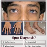
Answer is B. Homocystinuria
Key reasoning:
• Marfanoid habitus (tall stature + long limbs/arachnodactyly) + ectopia lentis (lens dislocation causing progressive visual blurring) occurs in both Marfan syndrome and classic homocystinuria.
• The decisive feature is the history of recurrent thrombotic episodes. Homocystinuria (cystathionine β-synthase deficiency) produces markedly elevated homocysteine, which is strongly prothrombotic. Marfan syndrome does not cause a primary thrombotic tendency.
• Lens dislocation is typically downward in homocystinuria (versus upward in Marfan), consistent with the appearance in the provided eye photo.
• Wilson disease presents with Kayser-Fleischer rings, hepatic/neurologic disease, and low ceruloplasmin — not this body habitus or thrombosis.
• Ehlers-Danlos syndromes feature joint hypermobility, skin fragility, and vascular fragility in certain types, but not ectopia lentis or this metabolic thrombotic picture.
This is a classic spot diagnosis distinguishing homocystinuria from Marfan syndrome.
31

Answer is B. Harlequin ichthyosis
The infant shows the classic appearance of severe congenital ichthyosis: thick, plate-like (armor-like) hyperkeratotic scales, taut shiny skin, and ectropion. This is the hallmark of harlequin ichthyosis, the most severe form of autosomal-recessive congenital ichthyosis.
• A (Lamellar ichthyosis) presents with milder, finer or larger plate-like scales and less extreme facial distortion.
• C (Ichthyosis vulgaris) is a milder, later-onset condition and does not look like this at birth.
• D (Epidermolytic ichthyosis) features blistering, erosions, and different scale morphology.
56

Answer is B. Octreotide with continued glucose monitoring
Glipizide is a sulfonylurea. In overdose it causes prolonged, recurrent hypoglycemia by stimulating pancreatic insulin release.
When hypoglycemia keeps recurring despite IV dextrose boluses and a glucose infusion, octreotide is the specific treatment of choice. It suppresses further insulin secretion, allowing glucose therapy to be more effective, and is given with ongoing glucose monitoring.
• A (IV glucagon alone) is suboptimal because glucagon can further stimulate insulin release in sulfonylurea overdose.
• C (IV sodium bicarbonate) has no role here.
• D (activated charcoal as sole treatment) may be considered early after ingestion but does not address established, recurrent hypoglycemia.
1
41

Answer is B. Potter sequence
This newborn shows classic Potter facies (flattened nose, low-set ears, micrognathia) plus clubfoot. These features result from prolonged oligohydramnios (oligohydramnios sequence), most often due to bilateral renal agenesis or severe urinary tract obstruction. The compression from low amniotic fluid produces the characteristic face and limb deformities.
• A (Cushing syndrome) does not present with this facial and limb pattern in a newborn.
• C (Turner syndrome) typically shows webbed neck, lymphedema, and other features, not Potter facies.
• D (Marfan syndrome) involves long limbs and connective-tissue findings that are not present at birth in this form.
42