

Intensivist and Cardiac Anesthesiologist. #POCUS education Tweets are my own
New York, USA
Joined August 2019
- Tweets155
- Following497
- Followers361
- Likes149






Stamatis Baronos retweeted

My Alma Mater @upbcolombia invited me to present a lecture as an alumnus. I shared my wisdom regarding ultrasonography. The current dean is an intensivist! I am inspired me to prepare my next Pulm/CCM Ground Rounds at my current institution - @BCMDeptMedicine, on April 9th. @SCCM


Stamatis Baronos retweeted

New collab with @openanesthesia, the biggest educational resource for anesthesiologists. Our POCUS videos now featured on openanesthesia.org.
@TheICUdoc @GabrielPradaMD
#POCUS #anesthesia #echofirst #ultrasound

@HopkinsSICUDrG @HopkinsACCM @GabrielPradaMD @Diaz_GomezJL @anesthap @Baylor
Watch the video at for COVID-19 sciencedirect.com/science/ar…
Thank you for the support! @Wilkinsonjonny

Now then...if you want to learn #POCUS and in a clear and fabulous, easy to follow format; check out this amazing website. I am in awe👏👏👏 @TheICUdoc ...well done!!!
theicudoc.com
#FOAMed #FOAMcc #echofirst #foamus #medtwitter

Here is the vaccine we are about to receive
This Post is from an account that no longer exists. Learn more
1

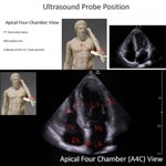
LA: left atrium
LV: left ventricle
IAS: interatrial septum
RA: right atrium
RV: right ventricle
TV: tricuspid valve
MV: mitral valve
IS: inferoseptal wall
AL: anterolateral wall
For full video on how to get the view check out youtu.be/mfUxeK86F6M
1
For full tutorials on basic and advanced views check out theicudoc.com
#echocardiography #echo #echofirst #cardiacsurgery #cardiacanesthesia #cardio #cardiacicu #cardiovascular #doctor #heart #health #icu #criticalcare #medicalstudent #POCUS #foamed #foamcc
3
Parasternal short axis view with focus on the Left Ventricle.
Excellent view to evaluate for regional wall motion abnormalities.

1
2
The basal and mid segments:
Anterior
Anterolateral
Inferolateral
Inferior
Inferoseptal
Anteroseptal
The apical segments:
Anterior
Lateral
Inferior
Septal
For a full video tutorial on how to get this view check out youtu.be/6tNfw1ggRQ0
1
1
False tendons are fibrous or fibromuscular bands attaching to LV walls or papillary muscles. Unlike true cordae, they do not attach to the mitral leaflets. False tendons are associated with LV hypertrophy and are better visualized when the LV is dilated.
How can you tell it's a false tendon?
1) Intracavitary course
2) Constant motion of the tendon throughout the cardiac cycle
3) Absence of regional wall motion abnormalities adjacent to the site of insertion
4) Presence of non-echogenic space on either side of the tendon
Don't confuse false tendons with accessory papillary muscles, subaortic membrane, abnormal leaflets, mass, thrombus or tumors.#echocardiography #echofirst
#cardio #cardiovascular #cardiology #cardiacsurgery #cardiologyfellow #cardiacanesthesia #cardiacicu #foamed #foam #foamcc