

Cardiac Surgeon | Assistant Professor | @TexasChildrens | Visual insights in surgery, cardiology & intensive care | Views my own
Joined July 2025
- Tweets2K
- Following106
- Followers8.1K
- Likes7.1K



Pinned Tweet
Left-to-Right vs Right-to-Left Shunting
👉Left-to-right shunts increase pulmonary blood flow and produce pulmonary overcirculation with volume loading of the involved chambers.
👉Right-to-left shunts bypass effective pulmonary oxygenation, causing systemic desaturation and reduced systemic oxygen delivery.
👉Shunt direction and magnitude depend on anatomy, pressure gradients, ventricular compliance, PVR/SVR balance, and pulmonary blood flow.


Fontan Circulation — Series: #1–10
👉Fontan circulation converts staged single-ventricle palliation into passive pulmonary blood flow without a subpulmonary ventricle.
👉Low PVR, adequate ventricular preload, and a favorable pressure gradient are essential despite systemic venous hypertension.
👉Venous congestion, abnormal shunting, and lymphatic dysfunction contribute to Fontan complications and failure.
↓ Full series below

10/10 | #10 Fontan Failure—Rescue Strategies and Takedown
👉Rescue strategies address PVR, PA obstruction, AV valve dysfunction, and Fontan pathway compromise, with takedown restoring Glenn physiology when necessary.
teapawt.org/WithAScalpel/status/21…

Fontan Circulation #10: Fontan Failure—Rescue Strategies and Takedown
👉Fontan failure involves elevated systemic venous pressure and impaired cardiac output, often requiring urgent hemodynamic intervention.
👉Rescue strategies include PVR reduction, PA reconstruction, AV valve repair, fenestration, and Fontan pathway revision.
👉When rescue strategies fail, Fontan takedown restores IVC return to the right atrium, reverting to Glenn physiology to relieve venous congestion.
95
9/10 | #9 High-Risk Fontan
👉Elevated PVR, ventricular dysfunction, AV valve regurgitation, and unfavorable vascular anatomy increase the risk of Fontan failure.
teapawt.org/WithAScalpel/status/21…
Fontan Circulation #9: High-Risk Fontan
👉Hemodynamic risk factors include elevated PVRi (>3 WU·m²), ventricular EDP >12 mmHg, AV valve regurgitation, and ventricular dysfunction.
👉Anatomic and surgical risk factors include prior PA or pulmonary venous interventions and an intact atrial septum.
👉Genetic syndromes, impaired ambulation, and respiratory support requirements further identify patients at high risk for Fontan completion.

7
8/10 | #8 Lymphatic Dysfunction in Fontan Circulation
👉Elevated CVP impairs lymphatic drainage, contributing to protein-losing enteropathy, plastic bronchitis, and chylothorax.
teapawt.org/WithAScalpel/status/21…
Fontan Circulation #8: Lymphatic Dysfunction in Fontan Circulation
👉Chronically elevated CVP increases lymphatic afterload, impairing drainage into the systemic venous circulation.
👉Elevated venous pressure and increased lymphatic flow promote lymphatic congestion, abnormal collateralization, and leakage.
👉Lymphatic dysfunction may manifest as protein-losing enteropathy (PLE), plastic bronchitis, or chylothorax.
11
7/10 | #7 Pulmonary Arteriovenous Malformations (AVMs)
👉Pulmonary AVMs cause intrapulmonary right-to-left shunting and systemic desaturation without directly decompressing the Fontan pathway.
teapawt.org/WithAScalpel/status/21…
Fontan Circulation #7: Pulmonary Arteriovenous Malformations (AVMs)
👉Pulmonary AVMs create intrapulmonary right-to-left shunting, bypassing the capillary bed and causing systemic desaturation.
👉Unlike Fontan fenestration, pulmonary AVMs cause desaturation without directly decompressing the Fontan venous pathway.
👉Pulmonary AVMs may develop after Glenn or Fontan palliation, particularly when hepatic venous effluent is excluded from a lung.
10
6/10 | #6 Systemic-to-Pulmonary Collaterals
👉Aortopulmonary collaterals bypass the cavopulmonary pathway, increasing pulmonary venous return and systemic ventricular volume loading.
teapawt.org/WithAScalpel/status/21…
Fontan Circulation #6: Systemic-to-Pulmonary Collaterals
👉Aortopulmonary collaterals commonly arise from the internal mammary, intercostal, bronchial, and thyrocervical arteries.
👉These vessels bypass the cavopulmonary pathway, delivering systemic arterial blood to the lungs and increasing pulmonary venous return.
👉Excessive collateral flow causes ventricular volume overload, elevated filling pressures, and potentially prolonged pleural effusions.
8
5/10 | #5 Fontan Fenestration
👉Fenestration provides a right-to-left shunt that reduces Fontan pressure and preserves cardiac output at the expense of systemic oxygen saturation.
teapawt.org/WithAScalpel/status/21…
Fontan Circulation #5: Fontan Fenestration
👉Fontan fenestration creates a right-to-left shunt from the Fontan pathway to the atrium, acting as a pressure-relief mechanism.
👉It reduces systemic venous pressure while preserving ventricular preload and cardiac output, particularly when PVR is elevated.
👉Fenestration may improve early postoperative hemodynamics and reduce venous congestion, at the cost of lower oxygen saturation.
6
4/10 | #4 Pressure Gradient and Systemic Venous Hypertension
👉Fontan circulation depends on a small pressure gradient from the systemic veins to the pulmonary venous atrium, sustained by systemic venous hypertension.
teapawt.org/WithAScalpel/status/21…
Fontan Circulation #4: Pressure Gradient and Systemic Venous Hypertension
👉Fontan flow relies on elevated systemic venous pressure (CVP often 12–18 mmHg) to drive blood through the pulmonary circuit.
👉The transpulmonary gradient (CVP − atrial pressure) is typically small (~3–6 mmHg), requiring low PVR for adequate flow.
👉Elevated PVR, atrial pressure, or ventricular EDP worsens venous congestion and compromises ventricular preload and cardiac output.
13
3/10 | #3 Transition from Glenn to Fontan Circulation
👉Fontan completion incorporates IVC return into the pulmonary circulation, improving systemic oxygenation while increasing venous pressure and congestion.
teapawt.org/WithAScalpel/status/21…
Fontan Circulation #3: Transition from Glenn to Fontan Circulation
👉Fontan completion incorporates IVC return into the Glenn circulation, establishing total cavopulmonary circulation.
👉Low PVR, low atrial pressure, and an adequate systemic venous-to-atrial pressure gradient are essential for maintaining cardiac output.
👉Fontan completion improves systemic oxygen saturation but increases systemic venous pressure, predisposing to venous congestion.
16
2/10 | #2 Fundamental Principles of Fontan Circulation
👉Fontan circulation relies on passive pulmonary blood flow driven by elevated systemic venous pressure, low PVR, and adequate ventricular preload.
teapawt.org/WithAScalpel/status/21…
Fontan Circulation #2: Fundamental Principles
👉Fontan completion directs SVC and IVC flow to the pulmonary arteries without a subpulmonary ventricle.
👉Pulmonary blood flow depends on the CVP–atrial pressure gradient, low PVR, and unobstructed pulmonary pathways.
👉Low PVR supports ventricular preload and output, while elevated CVP predisposes to venous congestion and lymphatic failure.
15
1/10 | #1 Staged Palliation for the Single Ventricle
👉Staged palliation progresses from parallel circulation to Glenn and Fontan physiology, progressively reducing ventricular volume overload.
teapawt.org/WithAScalpel/status/21…
Fontan Circulation #1: Staged Palliation for the Single Ventricle
👉Initial palliation balances Qp/Qs but often exposes the single ventricle to pulmonary flow–related volume overload.
👉The bidirectional Glenn directs SVC flow to the pulmonary arteries, reducing ventricular volume load while preserving IVC return.
👉Fontan completion routes both SVC and IVC flow to the lungs without a subpulmonary ventricle, requiring low PVR and adequate venous pressure.
23
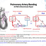
Pulmonary Artery Banding in VSD Physiology🫀
👉PA banding restricts pulmonary blood flow, reduces left ventricular volume overload, and lowers distal pulmonary artery pressure.


Fontan Circulation #10: Fontan Failure—Rescue Strategies and Takedown
👉Fontan failure involves elevated systemic venous pressure and impaired cardiac output, often requiring urgent hemodynamic intervention.
👉Rescue strategies include PVR reduction, PA reconstruction, AV valve repair, fenestration, and Fontan pathway revision.
👉When rescue strategies fail, Fontan takedown restores IVC return to the right atrium, reverting to Glenn physiology to relieve venous congestion.

ASD Patch Closure—Surgeon’s View🎯
👉A pericardial patch is secured to the atrial septum with a running suture to eliminate left-to-right shunting.

Answer Revealed! 🫀 — November 9 CMR Case
The patient had a large ASD with significant left to right shunting (Qp:Qs 5.9), severe mitral regurgitation (70% reg fraction), and moderate tricuspid regurgitation (30% reg fraction).
Read full case here 👉scmr.org/cases-of-scmr/numbe…
Fontan Circulation #9: High-Risk Fontan
👉Hemodynamic risk factors include elevated PVRi (>3 WU·m²), ventricular EDP >12 mmHg, AV valve regurgitation, and ventricular dysfunction.
👉Anatomic and surgical risk factors include prior PA or pulmonary venous interventions and an intact atrial septum.
👉Genetic syndromes, impaired ambulation, and respiratory support requirements further identify patients at high risk for Fontan completion.
Critical Closing Pressure—The Vascular Waterfall🏞️
👉Critical closing pressure (Pcc) creates a vascular waterfall, making microvascular perfusion dependent on the pressure gradient above Pcc rather than MAP alone.


🫀The vascular waterfall: why a higher MAP may not improve tissue perfusion
Mean arterial pressure is essential in shock, but it is not the pressure gradient that every organ actually experiences. A patient may reach the prescribed MAP while capillary flow remains inadequate. How can pressure improve without perfusion following?
Pinsky, Monge García and Dubin writesbabout critical closing pressure (Pcc), the pressure at which small, tone-dependent vessels cease conducting flow. Like a collapsible Starling resistor, the vascular segment creates an effective downstream pressure that may exceed venous pressure. Below this threshold, lowering venous pressure further does not increase flow: the vascular waterfall.
This changes the familiar interpretation of perfusion pressure. In the authors’ model, tissue perfusion pressure = MAP - Pcc, rather than simply MAP minus venous pressure. Their illustrative example is revealing: with MAP 75 mmHg and Pcc 35 mmHg, the effective gradient is only 40 mmHg. Pcc is not a fixed universal number; vascular beds differ in tone, metabolic demand and autoregulation.
In sepsis, loss of vasomotor tone may lower Pcc while simultaneously reducing MAP and disrupting local flow regulation. Conversely, norepinephrine may raise arterial pressure but also change vascular tone and Pcc. A higher MAP does not guarantee a larger effective gradient or better microcirculatory flow. The response depends on the patient's vascular state and the organ being considered.
Clinically, this provides a physiological explanation for discordance between macrocirculation and perfusion. Capillary refill, skin temperature, urine output, lactate trajectory and organ function remain essential alongside MAP and cardiac output. Pcc can be estimated from pressure-flow relationships, but global estimates cannot reliably describe every regional microvascular bed.
Reference 📚
Pinsky et al. 2024. Critical Care. doi.org/10.1186/s13054-024-0…
#Hemodynamics #Shock #Microcirculation #CriticalCare #ClinicalPhysiology

Fontan Circulation #8: Lymphatic Dysfunction in Fontan Circulation
👉Chronically elevated CVP increases lymphatic afterload, impairing drainage into the systemic venous circulation.
👉Elevated venous pressure and increased lymphatic flow promote lymphatic congestion, abnormal collateralization, and leakage.
👉Lymphatic dysfunction may manifest as protein-losing enteropathy (PLE), plastic bronchitis, or chylothorax.
