

ECG discussion page🫀 // everyone is welcome to take part im discussion 🗣️ or send their ECG-Cases 💬
Joined March 2022
- Tweets269
- Following104
- Followers3.1K
- Likes246
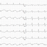

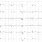
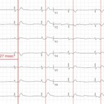
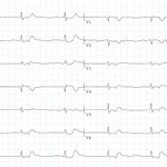
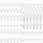

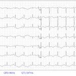
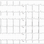
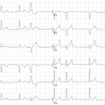

Something you don't see every day: HyperK+ in Pacemaker-ECG ⚡
@ecgrhythms @The_Nanashi_O @gerben_robbers
@BrooksWalsh @EPeeps_Bot @DidlakeDW @smithECGBlog

After 24h this poll is over. Unfortunately the majority of people was misled and chose the wrong answer. Based on this tracing, we cannot say, that there is complete AV-Block. We can only say, that there is AV-dissociation.
Why? Let's take a look! ⬇️⬇️⬇️


What is it and where does it come from? Your opinions? @ecgrhythms @The_Nanashi_O @gerben_robbers @BrooksWalsh @EPeeps_Bot @DidlakeDW #ECG


Typical CP in a 66y/o man radiating to the left arm for some hours, still present when ECG gets recorded, Troponin I at 1407ng/l. Your expertise?
HR 76bpm | PQ 186ms | QRS 136ms | QTc 468ms
@ecgrhythms @The_Nanashi_O @gerben_robbers @BrooksWalsh @EPeeps_Bot @DidlakeDW


This tracing from am man with waning CP was interpreted as PE due to "S1Q3T3". But is it really PE or rather something else?
#PMcardio @EPeeps_Bot #ECG
Poll⬇️


Pt 40a with worsening epigastric pain for hours. Then he called EMS, this ECG was recorded. Poll⬇️
Follow-Up will be provided in 24 hours.
@ecgrhythms @The_Nanashi_O @gerben_robbers @brun_dav @BrooksWalsh @EPeeps_Bot @DidlakeDW
#ECG


Do you recognize the subtle findings in this tracing from a 58y/o man with chest pain?
@EPeeps_Bot #ECG

Pt 55a with CP. V2 tells us the diagnosis, doesn't it?
@ecgrhythms @The_Nanashi_O @gerben_robbers @brun_dav @BrooksWalsh @EPeeps_Bot @DidlakeDW @smithECGBlog @ekgpress
#ECG

Patient got resuscitated for ~3 minutes. After the 2nd shock this ECG was recorded. BP 95/55, SpO2 83%. I think the diagnosis is quite obvious, but I like this ECG since it contains many different features of MI.
@EPeeps_Bot #ECG #EM

I'd like to continue the case: We interpreted the ECG as non-specific and decided to bring the Pt to the ED. After 15 minutes of transport, an alarm shows up at the monitor - HF is now ~ 30-40/min (see ECG). The patient is sweating, even more dizzyness but still denying any pain.

Man in his 80s collapses at home several times. No pain, only some dizziness. RR 110/72, SpO2 91%, BG 112mg/dl. What would you do and where would you bring him?
@ecgrhythms @The_Nanashi_O @gerben_robbers
@brun_dav @BrooksWalsh @EPeeps_Bot
@DidlakeDW @ahmad_70x
#EM #Cardiology

Man in his 80s collapses at home several times. No pain, only some dizziness. RR 110/72, SpO2 91%, BG 112mg/dl. What would you do and where would you bring him?
@ecgrhythms @The_Nanashi_O @gerben_robbers
@brun_dav @BrooksWalsh @EPeeps_Bot
@DidlakeDW @ahmad_70x
#EM #Cardiology