

We provide the most advanced #electrophysiology instrumentation to accelerate #drugdiscovery and advance basic #neuroscience research. #MEA #iPSC #Organoids
Switzerland
Joined June 2016
- Tweets1.3K
- Following891
- Followers1K
- Likes606




ISSCR 2026 Kyoto may have wrapped up, but our commitment to Amyotrophic lateral sclerosis (ALS) research continues.
While the conference brought together researchers to celebrate advances in iPSC science and explore new opportunities for disease modeling and regenerative medicine, one of our most meaningful moments happened beyond the scientific sessions.
🏃♂️On the crisp morning of Day 3, three members of our team @u_frey ,@engeleneo and @m_hruskaplochan took to the streets of Kyoto for a sunrise Run for ALS. We were delighted to be joined by other runners including Prof. Jun TAKAHASHI @juntakajun (@CiRA_KU_E Center for iPS Cell Research and Application (CiRA)) and Keith Alm (@ISSCR International Society for Stem Cell Research), sharing those early morning miles to raise awareness and show our support for people living with ALS.
ALS is a progressive neurodegenerative disease that affects motor neurons, gradually impairing the ability to move, speak, swallow, and breathe. Behind every diagnosis are people and families facing profound challenges, reminding us why continued scientific progress and collective action matter.
But our commitment extends beyond technology and the lab. Through initiatives like Run for ALS, we come together as colleagues and as part of a broader community united by the hope of making a difference.
💙 Every contribution counts. Support our initiative here: secure2.convio.net/alsa/site…
🙏🏻 A heartfelt thank you to everyone who joined us, cheered us on, or contributed to our fundraiser. What started in Zurich during NeuMoS continued through the streets of Kyoto, and our journey is far from over. Stay tuned for our next Run for ALS as we keep moving together for a cause that matters! 💙
#ALS #RunForALS #ALSResearch #ALSAwareness #ISSCR #MxWConnect


🫂One organoid? Two? Three? Which network can learn to discern one input from another? The more, the merrier.
In a new @CommsBio (@NaturePortfolio) paper, Chow et al. used MaxWell Biosystems’ MaxOne High-Density Microelectrode Array (#HDMEA) System to build and longitudinally stimulate solo, duo, and trio human cerebral #organoid networks, asking whether repeated electrical input could refine their ability to discriminate two different signals.
Using MaxOne+, the team could monitor all interconnected modules simultaneously while keeping their activity spatially resolved, enabling spike sorting and functional connectivity analysis across the network. These analyses revealed that, after two weeks of repeated input (aka training), only the three-organoid networks consistently improved stimulus discrimination, with the output organoid responding differently depending on which upstream organoid was activated. This functional refinement was also reflected in spontaneous activity, where network propagation became increasingly biased from the input organoids toward the output.
This work highlights how modular network architecture together with repeated external cues can drive stimulus-specific functional refinement in human brain organoid networks.
👉 Read the full publication here: mxwbio.com/resources/communi…
👏 Congratulations to Siu Yu A. Chow, Huaruo Hu LinkedIn, @Tomoyadu , Takuya Asakura, Sota Sugimura, and @YoshihoIkeuchi from @UTokyo_News_en, CNRS, and @SoftBank on their new publication!
#Connectoid #Organoids #FunctionalConnectivity #OrganoidIntelligence #Neuroengineering #Neurocomputing #ElectricalStimulation #iPSC #Neuroplasticity

💤 Can a human brain #assembloid reproduce the electrical signatures of #anaesthesia?
In a new British Journal of Anaesthesia paper, Toker et al. asked whether a minimal human cortical circuit, without the thalamus or other subcortical structures, could reproduce propofol-induced neural dynamics. Their cortical–ganglionic eminence assembloids provided this minimal circuit, combining excitatory and inhibitory neurons with propofol-sensitive GABAA receptors.
The team captured three hallmark electrophysiological features of anaesthetic states. Using MaxWell Biosystems’ MaxOne HD-MEA System and spike sorting, they observed an abrupt and sustained suppression of single-unit firing after propofol exposure, while complementary LFP recordings revealed a shift toward prominent slow/delta activity together with reduced signal complexity.
By reproducing key anaesthesia-like electrophysiological dynamics in a human in vitro model, this work opens new possibilities for mechanistic studies of anaesthetic action and future drug screening.
👉 Read the full publication: mxwbio.com/resources/british…
👏 Congratulations to Daniel Toker and all co-authors
#BrainOrganoids #HDMEA #MaxOne #iPSC #Neuroscience #GABA #Pharmacology

🧠 Two human dopaminergic neurospheroids. One axonal bridge. Can HD-MEAs reveal whether the two networks are actually interacting?
In a new Biofabrication (@IOPPublishing) paper, Ausilio et al. developed a microfluidic microphysiological system (MPS) in which two human iPSC-derived dopaminergic neurospheroids are kept spatially separated while axons grow through a defined microchannel, forming a stable inter-spheroid fascicle.
But does this morphological connection also translate into a functional one? In this case, yes.
Using MaxWell Biosystems’ MaxOne+High-Density Microelectrode Array (HD-MEA) System, the team followed the two neurospheroids longitudinally and could spatially resolve activity from each network while monitoring their maturation over time.
Going one step further, the authors separated local interactions within each spheroid from long-range interactions between them, revealing bidirectional lag-consistent activity across the connecting axonal tract, compatible with functional connectivitybetween the two neuronal modules.
This work brings 3D human dopaminergic circuitry models, microfluidic MPS, and HD-MEA electrophysiology together in one platform to investigate how spatially separated neuronal networks connect and communicate.
👉 Read the full publication here – mxwbio.com/resources/biofabr…
👏 Congratulations to Chiara Ausilio, Annachiara Scalzone, Valentina Mollo, and Paolo Antonio Netti from Istituto Italiano di Tecnologia (IIT) and Università degli Studi di Napoli Federico II on their new publication in Biofabrication / IOP Publishing.
#Connectoid #MicrophysiologicalSystems #MPS #HDMEA #Neurospheroids #Microfluidics #BrainOnChip #Neuroengineering #iPSC

It was a pleasure to welcome our partners to MaxWell Biosystems in Zurich, Switzerland, for our second annual IUK–Innosuisse consortium meeting!
A big thank you to Zameel Cader, Satyan Chintawar, Galbha Duggal, and Anna Underhill from Oxford StemTech, and Joseph Bateman and Harry Mayes from King’s College London, for the insightful discussions, productive exchanges, and great time together.
Together, we are developing a high-throughput drug testing platform combining patient-specific brain assembloids with high-density microelectrode array (HD-MEA) recordings, with the goal of advancing precision medicine for epilepsy.
It is exciting to see this collaboration progressing and to bring together complementary expertise in stem cell models, electrophysiology, and drug discovery to develop new approaches for patient-specific therapeutic testing.
We are grateful to Innosuisse and Innovate UK (IUK) for supporting this collaborative project.
🔗 Want to learn more about the project? Find more info here: mxwbio.com/collaborations#iu…
#Epilepsy #PrecisionMedicine #Organoids #NAMs #HighThroughput #HDMEA

🫀 Can engineered cardiomyocytes improve cardiac repair after myocardial infarction?
In a new Bioactive Materials paper published by KeAi Publishing, Lee et al. tackle one of the major challenges of cardiac cell therapy: the poor retention and survival of transplanted human pluripotent stem cell-derived cardiomyocytes (hPSC-CMs) in the injured heart.
The team developed a rapid surface-engineering strategy using lipid insertion and click chemistry to attach a collagen hybridizing peptide (CHP) to hPSC-CMs. By targeting denatured collagen in injured myocardium, CHP-modified cells showed improved adhesion and engraftment, supporting better cardiac repair after transplantation in a rat ischemia-reperfusion model.
But what about beating function?
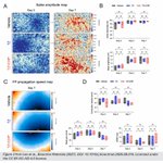
Using the MaxOne High-Density Microelectrode Array (HD-MEA) System, the team followed the cultures on days 2, 5, and 7 after modification. Active beating area, field-potential amplitude, beating rate, rhythm variability, and conduction velocity remained comparable across conditions, with propagation maps further confirming coordinated electrical conduction across the network — showing that the surface-engineered cardiomyocytes retained their electrophysiological phenotype.
Together, the study highlights how HD-MEA functional phenotyping can provide a quality-control step for engineered cell therapies, connecting cell-surface bioengineering with cardiac repair in vivo.
👉 Read the sull publication here:
mxwbio.com/resources/bioacti…
👏Congratulations to Donghyun Lee, Woo-Sup Sim, Jae-Hyun Park, Dong-Yeon Han, Soo-Jeong Kim, Jin Ryeol An, Mi Seon Seo, Jin-Ju Kim, Seo Young Cheon, Hyejin Hong, Seong Woo Choi, Kiwon Ban, Heebeom Koo, and Hun-Jun Park, from The Catholic University of Korea, @donggukuniv, and @CityUHKDG.
#CardiacResearch #Cardiomyocytes #StemCells #RegenerativeMedicine #HDMEA #Electrophysiology #CellTherapy #Bioengineering

Check out our new Cardiac Assay integrated in MaxLab Live! mxwbio.com/applications/card…
In 3D pharmacology, a robust baseline is only the starting point. The next question is: how does a compound reshape network function?
In our new joint application note with @InSphero, 3D InSight™ Human Neural Microtissues recorded on the MaxTwo 24-Well Plate showed reproducible spontaneous activity and synchronized network bursting across independent microtissues, establishing a consistent functional baseline for compound testing.
Following pharmacological modulation, HD-MEA recordings revealed distinct network responses to several neuroactive compounds. But the analysis goes beyond conventional network metrics. With the new burst-shape analysis in MaxLab Live, changes in the population bursts dynamics can be characterized in greater detail, from burst onset and peak activity to decay.
Together, reproducible 3D neural models, scalable HD-MEA recordings, and advanced network analysis bring greater granularity to the functional characterization of compound responses.
👉 Read the full application note: mxwbio.com/resources/mxw-ins…
#Pharmacology #Spheroids #Neuroscience #Electrophysiology #HDMEA

See what you can now quantify with MaxLab Live, from burst-shape dynamics to synchrony and network activity: mxwbio.com/updates/maxlab-li…
🧠 Advancing functional characterization of iPSC-based models of motor neuron disease
As the field advances from individual iPSC-derived neuronal populations toward increasingly sophisticated 2D and 3D models, new opportunities are emerging to capture the cellular complexity of diseases such as ALS, SMA, and inherited peripheral neuropathies. At the same time, questions around functional characterization, reproducibility, model quality, and translational relevance are becoming increasingly important.
⚡ How can electrophysiology contribute to this next generation of disease models?
At the iPSC Modeling in Neuromuscular Disorders (iMND) Meeting in Rotterdam, meet Dr. Marilina Douloudi and Dr. Tom Dufor, Field Application Scientists at MaxWell Biosystems, and discuss your models, functional phenotyping, and how HD-MEA technology can complement your iPSC workflow.
Do not miss their poster presentation:
📌 “Electrophysiological Profiling and Phenotypic Characterization in ALS and FTD Models Using High-Density Microelectrode Arrays”
Discover how HD-MEA can add a functional dimension to iPSC-based disease modeling by complementing molecular and cellular readouts with high-resolution measurements of neuronal activity, helping uncover functional alterations associated with neurodegenerative disease.
📍 Rotterdam, The Netherlands
📅 September 30 – October 2, 2026
#iMND2026 #DiseaseModeling #Electrophysiology #HDMEA #MxWConnect


🧫 From thalamic input to human cortical development
In a new paper published in @ScienceMagazine, Claudia V. Nguyen and colleagues use human thalamocortical assembloids, to investigate how signals from one brain region shape the development of another.
What makes this especially interesting is how early thalamic input influences the production of cortical excitatory neurons. The team identified NRXN1-mediated contacts between thalamic axons and cortical progenitor cells as a mechanism contributing to this process.
Using the MaxTwo HD-MEA system, the researchers characterized electrical activity in 12-week-old assembloids. Spike-waveform analysis revealed a shift towards broad-spiking waveforms in thalamocortical assembloids compared with cortex–cortex controls, adding an electrophysiological perspective to the study of how thalamic input shapes human cortical development (Sup. Fig. S6).
This creates an exciting experimental platform for studying how interactions between brain regions shape human cortical development, and how disease-associated NRXN1 mutations may disrupt this process.
Read the full publication here: mxwbio.com/resources/science…
🎉 Congratulations to Claudia V. Nguyen, Antoni Martija, Daniel C. Jaklic, Rista White, Marty G. Yang, Patricia R. Nano, Jose A. Soto, Jessenya Mil, Dakshesh Rana, Jacqueline M. Martin, Hunter E. Schweiger, Sebastian Hernandez, Elisa Fazzari, Yu Liu, Jack M. Parent, Mohammed A. Mostajo-Radji, Daniel H. Geschwind and Aparna Bhaduri, from @UCLA, @ucsc, @UMich and the VA Ann Arbor Healthcare System!
#HDMEA #Organoids #Assembloids #Neurodevelopment #Electrophysiology

👁️ Before the eyes even open, the retina is already “talking” to the brain. But what happens in neurodevelopmental disorders with visual impairment?
In a new Frontiers in Neuroscience paper (@FrontNeurosci), Gupta et al. investigate how loss of Ube3a, the gene underlying Angelman syndrome, affects spontaneous retinal activity during early development, a particularly interesting question given the visual abnormalities commonly associated with the disorder.
Before vision is fully established, developing retinas generate retinal waves, coordinated bursts of neuronal activity that travel across the tissue and help guide the wiring of the visual system.
Using the MaxOne High-Density Microelectrode Array (HD-MEA) System, the team recorded 32 ex vivo retinas from wild-type (WT) and Angelman-model mouse pups. Angelman-model retinas showed reduced retinal wave frequency and lower overall RGC activity, indicating less patterned activity during this critical developmental period.
But what about the waves that did occur? They were largely normal: wave propagation speed, duration, area, shape, local synchrony, and distance-dependent functional connectivity were comparable to WT controls. This suggests that Ube3a deficiency primarily disrupts the initiation of retinal waves rather than their ability to propagate across the retinal network.
This is a nice example of how the spatial and temporal resolution of MaxOne can move beyond detecting “more or less activity” and reveal which specific property of a developing neural circuit is altered in disease.
👉 Read the full publication here –
mxwbio.com/resources/frontie…
👏 Congratulations to Rishikesh Kumar Gupta, Alexander J. Fisch, Fernanda S. Orsi, and Alexandre Tiriac from Vanderbilt University (@VanderbiltU) and Vanderbilt University Medical Center (@VUMCDiscoveries) 🇺🇸
#HDMEA #Retina #Neurodevelopment #DiseaseModeling #Neuroscience
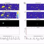
Supplementary Video S1 from Gupta et al., Frontiers in Neuroscience (2026). DOI: 10.3389/fnins.2026.1895084. Licensed under CC BY 4.0.
🚀 High content electrophysiology: from Earth to orbit.
We are excited to announce a new partnership between MaxWell Biosystems (@mxwbio) and Exobiosphere (@exobiosphere), bringing high-content electrophysiology to automated drug screening in microgravity.
By integrating our high-density microelectrode array (HD-MEA) technology into Exobiosphere’s Orbital High-Throughput Screener (OHTS), the collaboration aims to enable the functional characterization of neuronal and cardiac cells directly in orbit.
Why does this matter?
Microgravity offers a unique environment to investigate biological processes and drug responses. Adding high-content electrophysiology means going beyond observing how cells change in space to measuring how their function changes and how they respond to compounds.
The combination of automated screening and HD-MEA technology opens exciting opportunities to generate richer, functional insights for disease modeling and therapeutic discovery in microgravity.
We were delighted to share this next step together at NeuMoS 2026, and we are looking forward to seeing where this collaboration takes us. 🌍🛰️
👉 Read the full press release: mxwbio.com/updates/maxwell-b…
#MaxWellBiosystems #Exobiosphere #Microgravity #Electrophysiology #LifeSciences #SpaceResearch


🖐️🔥 Can engineered skin “feel” when it is touched, warmed, or cooled? And how those inputs are translated into electrical activity?
In a new Advanced Science (Wiley's @AdvPortfolio) paper, Bellantoni et al. present an innervated full-thickness human skin equivalent that turns physical stimuli into measurable neuronal responses. The model combines a differentiated human epidermis and dermis with Schwann cells and sensory neurons forming free nerve ending-like structures throughout the tissue.
Rather than evaluating sensory activation only through molecular or optical readouts, they interfaced the tissue with MaxWell Biosystems' MaxOne High-Density Microelectrode Array (HD-MEA) System to directly measure its electrical responses while applying physical stimuli.
And the skin responds.
Repeated mechanical indentation triggered increases in firing rate that became smaller with repeated touches, a potential signature of sensory adaptation. Thermal stimulation also modulated neuronal activity, with both warming and cooling producing a particularly clear increase in firing.
But can MaxOne distinguish mechanical from thermal stimuli? The authors found distinct electrical signatures in spike waveforms for mechanical versus thermal stimulation, demonstrating that the engineered tissue can encode information about stimulus modality.
This is a fascinating step toward experimentally controllable models of human somatosensation, with MaxOne at the functional interface between an engineered sensory tissue and the physical world.
👉 Read the full publication here – mxwbio.com/resources/advance…
👏 Congratulations to Daniele Bellantoni, Costantino Casale, Marika Sperduti, Nevio Luigi Tagliamonte, Giorgia Imparato, Loredana Zollo, and Paolo Netti from Università degli Studidi Napoli Federico II, Istituto Italiano di Tecnologia (@IITalk), and Università Campus Bio-Medico di Roma
#HDMEA #SensoryNeurons #Bioengineering #Somatosensation #Neuroengineering

👁️ Exploring retinal function together!
We are excited to join Helsinki Vision 2026 – Visual Neuroscience in Health and Disease.
📍 Helsinki, Finland
📅 September 16–18, 2026
At MaxWell Biosystems, we are helping researchers uncover new insights into retinal function with high-density microelectrode array (HD-MEA) technology.
With MaxOne, retinal explants can be investigated at high spatial and temporal resolution—from capturing retinal ganglion cell activity across the tissue to resolving signals at the single-cell and subcellular level, investigating axonal signal propagation, and characterizing responses to controlled light stimulation.
👋 Make sure to stop by our booth and meet Dr. Elisa Toscano, Field Application Scientist at MaxWell Biosystems! Whether you are working with retinal explants, investigating visual circuits, or curious about what HD-MEA technology could bring to your research, Elisa will be there to discuss your applications and show you MaxOne in action.
🔬 And there’s more on September 17! Don’t miss our Technology Spotlight during the Hands-On Workshop, followed by the opportunity to experience the technology firsthand at our dedicated demo station.
We are also delighted to have Dr. Annalisa Bucci, MaxOne user and recent Swiss OphthAWARD 2026 winner, participating in the workshop and bringing a researcher’s perspective on the technology.
Looking forward to connecting with the vision research community in Helsinki! 👁️⚡
#Retina #VisionResearch #Electrophysiology #HDMEA #Neuroscience #MxWConnect


🧠 Do you remember neurons playing Pong? Was getting better at it simply a matter of becoming “more connected”?
In a new Communications Biology paper (@NaturePortfolio and @SpringerNature), Khajehnejad et al. introduce TAVRNN, a graph-based machine-learning framework that tracks how neuronal activity and functional connectivity reorganize over time, and how this relates to behaviour.
The framework was validated on three electrophysiological datasets: rat hippocampus during navigation, primate somatosensory cortex during reaching, and DishBrain, where living neuronal networks interact with Pong through closed-loop stimulation.
The authors revisited the original DishBrain dataset, recorded using the MaxOne High-Density Microelectrode Array (HD-MEA) System: 24 cultures and 437 Pong Gameplay and Rest sessions.
Did neural connectivity simply increase during Gameplay? Conventional connectivity metrics could distinguish Gameplay from Rest, but were not sufficient to separate the best from the worst-performing Gameplay periods. So, what changed during better Gameplay? TAVRNN revealed that during high performance, sensory- and motor-associated channels became increasingly organized into structured functional clusters. During poorer performance, this organization was weaker and less clearly defined.
In other words, successful behaviour was associated not simply with “more connectivity”, but with dynamic and coordinated sensory–motor network organization over time.
👉 Read the full publication here – mxwbio.com/resources/communi…
👏 Congratulations to Moein Khajehnejad, Forough Habibollahi, Ahmad Khajehnejad, Chris French, Brett J. Kagan, and Adeel Razi from the Turner Institute for Brain and Mental Health, @MonashUni, @CorticalLabs , @UBC, @MelbourneOf, @ucl, and CIFAR on this work published in Communications Biology, part of @NaturePortfolio and @SpringerNature.

🧠 Bringing next-generation functional insights to human neural models.
From iPSC-derived neurons to increasingly complex organoids and assembloids, advanced human neural models are opening new possibilities for studying development, disease, and therapeutic response. Understanding their functional organization and network dynamics is key to unlocking their full potential.
At the 14th GSCN Conference in Berlin, our Field Application Scientist Dr. Elisa Toscano will showcase how high-density microelectrode array (HD-MEA) technology enables high-resolution functional characterization of human neural organoids and assembloids.
📌 Poster #53
“Next-Generation Electrophysiology for Functional Characterization of Human Neural Organoids and Assembloids”
📅 September 9 | 16:30–18:30
📍 Attending GSCN 2026? Stop by Elisa’s poster or connect with her throughout the conference, September 9–11.
Looking forward to connecting with the stem cell and organoid community in Berlin! 🧠⚡
#GSCN2026 #Organoids #Neuroscience #HDMEA #MxWConnect


🧠 From axon guidance and synapse formation to circuit function: how do neuronal connections give rise to coordinated network activity?
MaxWell Biosystems is joining the 15th Molecular Mechanisms of Neuronal Connectivity meeting at Cold Spring Harbor Laboratory, bringing together researchers exploring how neural connections form, develop, adapt, and regenerate.
📍 September 8–11 | Cold Spring Harbor Laboratory, NY
⚡ Interested in following neuronal activity and connectivity in unprecedented detail?
Our Dr. Carlos Sanchez Priego and Dr. @Fraser_mccready will be there to discuss how high-density microelectrode array (HD-MEA) technology can add a functional dimension to the study of neuronal connectivity, from mapping activity across neural networks to investigating axonal propagation and circuit dynamics at high spatial and temporal resolution.
We look forward to connecting with the neuronal connectivity community at #CSHL!
#cshlneurocon #Neuroscience #Electrophysiology #HDMEA #MxWConnect


Keep your organoid stable during recording, and ready for what comes next.
For acute organoid recordings, reliable positioning over the electrodes is key to capturing functional activity, while keeping sample handling as gentle as possible.
The MaxOne Organoid Holder uses a membrane-based design to gently keep the organoid positioned on the MaxOne+ HD-MEA Chip array area throughout recording, minimizing perturbation to the sample.
And the experiment does not have to end when the recording does. The organoid can be recovered afterward for follow-up experiments, further characterization, or complementary analyses, helping you to get more insights from every sample.
🔹 Gentle, reliable positioning during acute recordings.
🔹 Minimal perturbation to the sample.
🔹 Recover your organoid for what comes next.
Record the function. Preserve the sample. Continue the story.
👉 Discover the MaxOne Organoid Holder: mxwbio.com/products/maxone#M…
#Organoids #Electrophysiology #HDMEA #Neuroscience #3DModels