PhD Student @unibern IPS | she/her Studying stomatal development in grasses 🌾 Human Rights | Feminism | Climate Justice |
- Tweets126
- Following522
- Followers269
- Likes3.8K


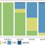




ALT Figure 1: (A) Schematic displaying stomatal development in wild type (WT). Open questions regarding the roles and mechanisms of BdMUTE are indicated. (B) In bdmute mutants, no subsidiary mother cell (SMC) is established, and no subsidiary cell (SC) division occurs. Guard mother cells (GMCs) mostly divide normally and the guard cell (GC) pair forms a pore and matures into functional complexes (upper GCs, dark green). However, ∼25% of the GMCs show aborted complexes with oblique divisions (lower GCs, gray). (C) Different stomatal morphologies found in WT (2 SCs), and in sid (1 SC, 0 SC and arrested phenotype). The images depicting the 1 SC and 0 SC phenotype stem from the weak rescue line M3GM. Midplane confocal images of fixed, cleared and Direct Red 23-stained mature stomata. Scale bars: 10 μm.


ALT Schematic displaying stomatal development in wild type (WT). Stomatal development starts with guard mother cell (GMC; blue) formation. BdMUTE is expressed in the GMC (light blue nucleus) and moves into the lateral cells (light yellow nucleus) to establish subsidiary mother cells (SMCs). SMCs then polarize (yellow gradient) and divide asymmetrically to form lateral SCs (yellow). Finally, the GMC divides symmetrically to generate two GCs (light green), which form a pore and mature into the characteristic dumbbell shape (dark green). Open questions regarding BdMUTE’s roles and mechanisms are indicated.

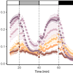
ALT gSW of WT, the four sid/bdmute-1;BdMUTEp:3xGFP-BdMUTE (M3GM) lines (++, +, - and - -) and sid/bdmute-1 under changing light conditions sowing a gradual rescue of the sid phenotype.