

Radiologist. Trauma and Emergency Radiology. One view is NO view. #radtwitter | #FOAMrad | #radres | #radEd| #radiology | #EmergencyRad
Muscat, Oman
Joined March 2013
- Tweets10.6K
- Following4.4K
- Followers19.7K
- Likes14.3K





Sameer Raniga retweeted

SUPRASPINATUS TEARS: UNDERSTANDING THE PATTERNS
Not all rotator cuff tears are the same. Understanding the relationship between the tear and the tendon surfaces is essential for accurate musculoskeletal ultrasound assessment.
This educational series illustrates the different patterns of rotator cuff tearing:
1. Articular-sided partial-thickness tear
2. Bursal-sided partial-thickness tear
3. Intrasubstance (interstitial) tear
4. Full-thickness tear
A systematic assessment of the articular surface, bursal surface, tendon substance, and extent of fiber disruption helps characterize these abnormalities and supports structured ultrasound reporting.
📷 For educational purposes: These illustrations are designed to facilitate understanding of supraspinatus tear morphology and ultrasound anatomy among radiologists, sonologists, and musculoskeletal imaging enthusiasts.
P.S. AI disclosure: All images in this series are AI-generated educational illustrations. They are schematic representations intended to explain tear patterns and are not actual ultrasound images or patient cases. They may not reproduce all anatomical and sonographic details accurately. Clinical interpretation should always be based on real-time ultrasound findings and appropriate clinical correlation.
#MusculoskeletalUltrasound #MSKUltrasound #Supraspinatus #RotatorCuff #RotatorCuffTear #ShoulderUltrasound #MSKRadiology #MusculoskeletalRadiology #UltrasoundEducation #RadiologyEducation #Sonography #SportsImaging #AIinMedicalEducation #MedicalImaging


From today's reporting list: a fracture in AVN that doesn't look like a crescent sign.
This is humeral head avascular necrosis in a patient with sickle cell disease.
Radiographs shows a geographic lesion in the superomedial humeral head with a curvilinear sclerotic margin. Along that margin runs a linear lucency, following the edge of the infarct rather than the articular surface.
MRI (coronal fat-suppressed T2): a thin fluid-bright cleft (arrow) at the interface between necrotic and reparative bone. It runs obliquely across the head, with marrow edema in the adjacent bone.
This is not the classic crescent sign, which sits just under the subchondral plate and parallels the joint surface. But it is still a fracture of the necrotic segment, just in a different plane.
Why does it happen here?
The necrotic segment can fail in two places:
1⃣ Subchondral, just under the plate → the crescent sign
2⃣ At the necrotic–reparative interface, where dead bone meets the stiff, thickened trabeculae of the repair zone → this case
Why it matters:
▪️ ARCO 2019 stage III includes fracture through the necrotic portion, not only the crescent sign
▪️ Call it a fracture of the necrotic segment, not a "subchondral insufficiency fracture." SIF is a separate entity where the fracture is primary.
Features that favour a fracture over the double-line granulation band:
✔️ Thin, sharp, fluid signal on all sequences, not enhancing
✔️ Marrow edema in the adjacent viable bone
✔️ Watch for contour step-off; CT is the most sensitive test
Take-home: in AVN, look for the fracture along the infarct margin, not only under the articular surface.
—Pearls, pitfalls and wisdom from my reporting list


A “broken heart” 💔 and a “lost ice cream cone” 🍦 !
Two classic CT signs of traumatic incudomalleolar joint dislocation.
Axial CT: Ice cream cone sign
The malleus head forms the ice cream, while the incus forms the cone. Loss of this configuration suggests dislocation.
Coronal CT: Broken heart sign
Disruption of the normal heart-shaped incudomalleolar articulation.
Take-home message:
In temporal bone trauma, don’t just look for fractures. Check the ossicular chain in both planes. Sometimes, a broken heart and a missing ice cream cone explain the hearing loss.
#EmergencyRadiology #Neuroradiology #RadiologyTeaching
—Teaching Pearl from My On-Call Cases | Temporal Bone Trauma


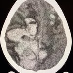
A brain intraparenchymal hematoma with a fluid-fluid or hematocrit level suggests impaired or delayed clot formation.
Think of:
• Anticoagulation
• Thrombocytopenia
• Coagulopathy
• Hematological disorders
• hyperacute or ongoing hemorrhage
—My on-call reporting list


Previous decompressive craniectomy, now with a markedly sunken scalp flap and inward deformation of the underlying brain.
One useful reporting point:
A sunken flap is an imaging finding. Sinking skin flap syndrome, or syndrome of the trephined, is a clinical-radiological diagnosis.
So if the clinical deterioration is not known, avoid diagnosing the syndrome from CT alone.
Describe what you actually see:
• sunken scalp flap
• inward deformation of the underlying brain
• midline shift
• ventricular changes
• paradoxical herniation, if present
The syndrome is considered when these findings are associated with neurological symptoms such as headache, weakness, cognitive slowing, gait disturbance, reduced consciousness, or failure to progress neurologically.
Cranioplasty may improve symptoms by restoring more normal pressure relationships, CSF dynamics and cerebral perfusion.
A useful distinction for trainees:
Report the morphology on imaging. Call it a syndrome only when the clinical picture supports it.
—Pearls, pitfalls and tips from my reporting list




Classic Signs in Radiology | Bone-within-Bone appearance
This radiograph from a patient with sickle cell disease shows the characteristic bone-within-bone appearance.
A smaller, denser outline appears within the normal contour of the bone: the bone-within-bone sign.
But what actually produces the “bone within the bone”?
In sickle cell disease: Chronic medullary infarcts develop linear or serpiginous sclerosis, which may run parallel to the cortex and outline the medullary cavity.
—Classic signs from my reporting list


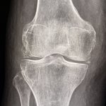
Chondrocalcinosis. Where to look?
Look at the menisci. Look at the articular cartilage, particularly along the femoral condyles. Here, it is nicely seen posteriorly.
Then look for effusion and soft tissue edema. Both are striking here, while degenerative change is minimal.
Large effusion + little OA + chondrocalcinosis = think acute CPPD.
But don’t forget infection.
Aspirate the joint. Exclude infection. Look for CPP crystals.
—pearls, pitfalls and wisdom from my reporting list


Sameer Raniga retweeted
Most people (including plenty of radiologists) misunderstand "resolution" in medical imaging, and the recent @midjourney debate hasn't helped. What resolution actually is, why your scanner's limit is set on day one, and the limit set by a Swedish-American named Nyquist 🧵1/10.

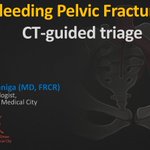
Excited to be speaking at the Radiopaedia 2026 Annual Meeting.
My session is on “Pelvic Vascular Trauma imaging”, where I'll share a practical CT approach to answering the questions that matter most in the trauma bay:
• Where is the bleeding?
• Which vessel is most likely injured?
• What should happen next?
The session introduces a simple 3B approach that integrates Blood, Bones, and Blush to localize hemorrhage, predict the injured vascular territory, and guide management.
This year's meeting brings together 56 international speakers, multiple parallel sessions, hands-on workshops, anatomy reviews, panel discussions, rPosters, and more than 14,000 delegates from around the world.
Looking forward to learning alongside colleagues from around the world.
Dates: 16 to 21 July 2026
Online and worldwide with free access in 125 countries.
Register here: radiopaedia.org/courses/radi…
Here's a sneak peek at a few slides from my talk.
@Radiopaedia
@thexraydoctor
@DrAndrewDixon
@teachplaygrub
@daniel_gewolb


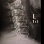
Don’t overcall anterior cervical fragment an "osteophyte fracture."
This lateral cervical radiograph illustrates a common reporting pitfall.
At first glance, the anterior triangular ossicles could be mistaken for fractured osteophytes.
However,
• They are well corticated.
• They have a typical location within the anterior annulus/anterior longitudinal ligament.
• There are no accompanying signs of acute injury.
These are degenerative intercalary (intercalated) ossicles, often related to annular calcification or chronic ossification, not acute fractures.
The diagnosis of an acute fracture should be supported by evidence of acute injury, such as cortical disruption (donor site from vertebra), prevertebral soft tissue swelling, associated vertebral or disc injury, or marrow edema on MRI.

Quiz: Linear radiopaque material tracking along both sides of the skull on CT. What is it?
Answer: Post-procedural changes from middle meningeal artery embolization for chronic subdural hematoma.
Why we do it
•Chronic subdural hematoma is not just “old blood.”
•It is a biologically active membrane disease.
•Outer neomembrane develops along the dura.
•It is vascular, fragile, and fed largely by branches of the middle meningeal artery.
•Recurrent microbleeds and exudation maintain or enlarge the collection.
•Embolization shuts this supply → less rebleeding → lower recurrence.
Where it fits clinically
•Adjunct to burr-hole evacuation to reduce recurrence
•Standalone in selected patients (mild symptoms, high surgical risk)
•Recurrent or bilateral chronic SDH
•Increasing use with growing evidence, but still evolving practice patterns
—Pearls, pitfalls and wisdom from my reporting list


In thalassemia major, SWI can show prominent susceptibility within the choroid plexus, reflecting iron deposition.
Seen at:
• Lateral ventricles
• Roof of 3rd ventricle
• Roof of 4th ventricle and foramen of Luschka
Also note:
• Diploic space widening
• Diffuse low T1 marrow signal (marrow hyperplasia)
Correlation with systemic iron overload is essential.
—Pearls, pitfalls and wisdom from my reporting list


“Hair-on-end” skull: X-ray and CT correlation
X-ray
•Widened diploic space
•Vertical radiating trabeculae
•“Hair-on-end” appearance
CT sagittal:
•Expanded diploic space
•Coarse, thickened trabeculae
•Perpendicular orientation
•Intervening low-density marrow
CT axial: expanded marrow spaces with intervening thickened trabeculae looks like a sponge 🧽
Think
•Marrow hyperplasia from chronic anemia
•Most often thalassemia major
•Less likely due to sickle cell and other chronic hemolytic states
—Classic signs from my reporting list


Papilledema on CT.
Look for subtle posterior globe flattening.
In the right context, think raised intracranial pressure.
And don’t stop there.
Ask why.
One important cause you don’t want to miss is cerebral venous sinus thrombosis (CVST). Especially when the brain parenchyma still looks deceptively normal.
—Pearls, pitfalls and wisdom from my reporting list


AP chest:
Right heart border absent.
No part of the heart lies to the right of the spine.
Middle lobe disease or chest wall deformity?
Lateral view:
Depressed sternum.
Diagnosis: Pectus excavatum.
Wisdom: Absent right heart border with leftward shift, think chest wall before lung.
—Nothing fancy today

