
I send you forth as sheep in the midst of wolves: be ye therefore wise as serpents, and harmless as doves.
Joined September 2024
- Tweets1.1K
- Following183
- Followers185
- Likes2.5K



billwilliam retweeted

Non rien...
Vous vous réveillez quand ?
La Spike dans les cellules foetales ? Ça donne quoi ?
Ben... on sait pas 🤷♀️🤦♀️🥴🤪🤬
Soyons clair... on sait que les cellules foetales responsables du développement du cerveau sont susceptibles à la Spike.
Je reprends les mots de @ZdenekVrozina :
"Une équipe italienne a prélevé des échantillons de tissus chez 17 femmes infectées qui ont interrompu leur grossesse entre les semaines 8 et 12. L’ARN viral a été détecté dans environ 5 FOETUS sur 17.
Toutes les femmes étaient ASYMPTOMATIQUES et AUCUNE n’avait de virus dans le sang."
Mais les conséquences ? Non mesurées... ben oui... pourquoi faire ?
Bon... les chercheurs disent quand même que, vu les impacts du SARS-CoV-2 (et de sa Spike), documentés, ce serait bien qu'on regarde...
Oh ! Si peu... parce que les effets subcliniques ou différés possibles (inflammation bas grade, mécanismes amyloïdes, altérations de substance blanche) : même sans symptômes maternels évidents, une exposition prénatale pourrait contribuer à des impacts neurodéveloppementaux discrets à long terme. Hein...
Alors, on va me dire que je râle tout le temps... mais flûte... ça sort dans la littérature. Et pas un peu... et ça roupille...

billwilliam retweeted

A PLA-linked military lab hand-carried undeclared coronavirus material to an NIH-funded US lab in 2018, wrapped in paper towels. CBP caught it. The FBI’s WMD unit took it. Same courier caught again in 2021 with 132 more vials. Rand Paul’s docs.
@SenRandPaul


billwilliam retweeted

According to Dept of Energy's scientists at National Labs, if SARS CoV2 came from animals it would require THREE intermediate hosts.
ZERO have been found.
They say a lab origin is much simpler to explain.
@julianbarnes @johnstravis @martinenserink @jeffmervis @ScienceMagazine

billwilliam retweeted

Eight MERS cases reported in Saudi Arabia.
6 of them report no known camel exposure.
Why SARS-CoV-2 spike protein drives cancer angiogenesis
teapawt.org/billwilliam321/status/…

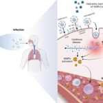
Metalloproteinases (MMP) are activated by inflammatory cytokines, Reactive oxygen species (ROS), and angiotensin II. [1]
The spike protein’s interactions with TLR2/4 and integrin probably drive inflammation. The spike protein also competitively inhibits ACE2 function, and the resultant buildup of angiotensin II leads to inflammation and production of ROS.
Excess MMP activity drives carcinogenesis.
By the same token, these stimuli also drive cancer angiogenesis. Cytokine storm, VEGF (from hypoxia and HIF activation), endothelial cell (EC) dysfunction, dysregulated Renin-angiotensin-aldosterone system (RAAS), and thrombosis contribute to angiogenesis. [2]
Spike protein promotes production of inflammatory cytokines (for example IL-6) by interaction with Toll-like receptors.
Hypoxia-Inducible Factor (HIF-1) can be activated by either hypoxia or hypoxia-independent mechanism.
Phosphatidylinositol 3-kinase (PI3K), which is downstream of TLR activation, also enhances HIF translation and subsequent VEGF production in hypoxia-independent mechanism. [3]
“Hormones such as angiotensin II and platelet-derived growth factor stimulate the HIF pathway by increasing HIF-1α protein levels via production of reactive oxygen species (ROS) within the cell.” [3] Excess angiotensin II also stimulates HIF pathway and VEGF production.
References:
1. Salomão R, Assis V, de Sousa Neto IV, Petriz B, Babault N, Durigan JLQ, de Cássia Marqueti R. Involvement of Matrix Metalloproteinases in COVID-19: Molecular Targets, Mechanisms, and Insights for Therapeutic Interventions. Biology (Basel). 2023 Jun 10;12(6):843. doi: 10.3390/biology12060843. PMID: 37372128; PMCID: PMC10295079.
pubmed.ncbi.nlm.nih.gov/3737…
2. Madureira G, Soares R. The misunderstood link between SARS-CoV-2 and angiogenesis. A narrative review. Pulmonology. 2023 Jul-Aug;29(4):323-331. doi: 10.1016/j.pulmoe.2021.08.004. Epub 2021 Aug 27. PMID: 34593362; PMCID: PMC8390375.
pubmed.ncbi.nlm.nih.gov/3459…
3. Ziello JE, Jovin IS, Huang Y. Hypoxia-Inducible Factor (HIF)-1 regulatory pathway and its potential for therapeutic intervention in malignancy and ischemia. Yale J Biol Med. 2007 Jun;80(2):51-60. PMID: 18160990; PMCID: PMC2140184.
pubmed.ncbi.nlm.nih.gov/1816…


billwilliam retweeted

2021年、「HIVの脳障害のメカニズムがコロナの神経症状を理解する枠組みになる」と提案された。5年分の臨床・病理・実験データが蓄積した今、その枠組みを見直す時が来た。最新の証拠は、コロナの神経症状が「ウイルス本体の直接感染」よりも「宿主の免疫反応」を主役とする方向へと重点を移した。持続的な免疫活性化、血管内皮の機能障害、グリア細胞の活性化、血栓炎症、そして神経免疫軸の破綻が、脳を壊す真のメカニズムであることが分かってきたのである。同時に、ウイルスの抗原(部品)が組織に残存していることや、一部の患者で自己抗体が検出されることも明らかになり、患者ごとに犯人が違うという生物学的な不均一性が浮き彫りになった。HIVの脳障害もまた、ウイルスの直接破壊ではなく免疫を介した間接的な破壊が主役である。コロナも同じ方向性を示しており、HIVの…
…
angama.bearblog.dev/hiv5/

billwilliam retweeted

ロシア国境に近い中国内モンゴルのフルンボイル市、ペスト危険注意報(中央日報日本語版)
news.yahoo.co.jp/articles/c9…

billwilliam retweeted
SARS-CoV-2 spike protein induces metalloprotease release by several mechanisms:
1. The spike protein binds Toll-like receptors (TLR2/4) and triggers hyperinflammation. Inflammatory pathways (IL-6 or NF-κB) probably elicit production of metalloproteases such as MMP-8 or MMP-9.
pubmed.ncbi.nlm.nih.gov/4058…
Besides spike protein, ORF8 also triggers hyperinflammation by mimicking IL-17.
Nucleocapsid protein, which is actively transported out of cell, activates complement system to attack health tissue—this is probably inflammatory as well.
2. ACE2 normally converts angiotensin II into angiotensin (1-7). Binding of spike protein disrupts ACE2 function and causes buildup of angiotensin II, which triggers inflammatory pathway.
pubmed.ncbi.nlm.nih.gov/4204…
3. RGD motif allows the spike protein to bind cell surface integrin. Spike protein binding to integrin activates mechano-sensitive Calcium channel TRPV4. High influx of Calcium ions leads to activation of pro-inflammatory pathway such as NF-κB and may induce release of metalloproteases.
Calcium influx also destabilizes tight junction in endothelium.
“Spike-ACE2 interaction activates ARF6 and NFκB signaling and promotes von Willibrand factor (vWF) release and down regulation and redistribution of key endothelial junctional proteins, including adherens junctions, junctional adhesion molecules (JAMs), and connexins. This disruption results in increased vascular permeability and a heightened risk of thrombosis.”
pubmed.ncbi.nlm.nih.gov/4167…


billwilliam retweeted

Russia’s plague appears to be spreading deep into China. We need to quarantine and test all arrivals from China and Russia.

On October 14, in response to the plague outbreak in Russia, the Centers for Disease Control and Prevention in Chenzhou and Yongzhou, Hunan Province, issued emergency alerts for plague prevention on the same day. Given that Hunan Province is thousands of kilometers from the Russian border, the occurrence of a plague outbreak at such a distance indicates that the Russian plague epidemic has already spread throughout China.
billwilliam retweeted
Deadly Pneumonic Plague Spreading? Hong Kong Alert, Rumors of Deaths Among China’s Patients & Medics
youtu.be/zwUfbGQRJuo?si=wOaJ…

billwilliam retweeted

Karolinska Institutet researchers found that SARS-CoV-2 infection accelerated lung tumor growth in mice by triggering low oxygen, inflammation, and VEGF-driven blood vessel formation.
Blocking VEGF significantly reduced tumor growth.
nature.com/articles/s41392-0…

billwilliam retweeted
コロナ感染で肺が低酸素になると、血管を作る信号物質が180倍に跳ね上がる。この異常な血管新生が、何年も眠っていた肺がん細胞を目覚めさせ、成長を開始させる——カロリンスカ研究所のチームがこのメカニズムを解明。マウス実験では、コロナ感染マウスの100%に肺がんができたのに対し、非感染マウスは28%だった。がん細胞の注入が少量でも、信号物質が多ければ腫瘍が形成される。つまり信号物質の量が「血管新生のスイッチ」をONにするかどうかを決める。
このスイッチを抗VEGF抗体でブロックすると、腫瘍の成長は止まり、多くが休眠状態のまま残った。ヒトのコロナ剖検肺でも、低酸素マーカーが9倍、VEGFと血管密度が有意に増加しており、マウスと同…
…
angama.bearblog.dev/on/
Why SARS-CoV-2 spike elicits autoantibodies against beta-adrenergic receptors and GPCR like MAS1
teapawt.org/billwilliam321/status/…
That’s a possibility.
A virus triggers auto-antibodies by three mechanisms:
1) Anti-idiotype network
2) molecular mimicry (similar motifs)
3) tissue damage and release of self antigen
Autoantibodies against GPCR may be raised by anti-idiotype mechanism. The word “idiotype” refers to the antigen-binding part of an antibody. The viral spike protein elicits primary antibodies (Ab1), and if the level of Ab1 is too high, the body also produces secondary antibodies (Ab2) attacking the primary antibodies at the idiotype site. Ab1 is complementary to epitope on the spike protein, while Ab1 is also complementary to Ab2. Therefore, Ab2 may mimic the spike protein in shape, and Ab2 may behave like the spike protein to interact with human receptors.
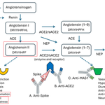
“Collins et al. [108] showed that sequential SARS-CoV-2 mRNA vaccination of hACE2 transgenic mice can induce both anti-S and anti-ACE2 antibody responses, and the latter was gradually amplified with each boost.” The spike protein raises antibodies against both the spike protein and the ACE2 receptor. Given that SARS-CoV-2 spike RBD binds β-adrenergic receptors with high affinity, the spike protein probably elicits autoantibodies against β-adrenergic receptors by anti-idiotype mechanism.
In addition, some of the spike motifs mimic human proteins in the blood coagulation pathway, thus inducing production of pro-thrombotic autoantibodies. For example, TQLPP motif on spike mimics thrombopoietin and may induce thrombocytopenia. ELDKY motif on spike mimics protein kinase G1 (involved in platelet activation).
Spike RBD may share some degree of similarity with angiotensin II, since both function as ligands for ACE2 receptor. Antibody targeting spike RBD may cross-interact with angiotensin II, leading to undesired vascular pathology such as hypertension.
Some parts of spike protein mimic ACE2. “Segments of the SARS-CoV-2 S protein (residues 49–53, 226–230, 418–428, 529–536, 744–774, and 977–980) share structural and chemical similarities with some segments of the ACE2 receptor (residues 137–141, 335–345, 350–352, 463–471, and 545–573).”
ACE2 converts angiotensin II (DRVYIHPF) into angiotensin (1-7) (DRVYIHP). A G-protein-coupled receptor MAS1 binds angiotensin (1-7) to mediate vasodilation. Because the two receptors use very similar ligands, the ligand-binding sites on these receptors are also similar. Anti-ACE2 antibody may cross-react with MAS1.
Reference:
Bellavite P, Di Fede G, Mantovani M, Zanolin E. Autoimmune Features of Post-COVID-19 Vaccination Syndrome and Their Impacts on the Renin-Angiotensin System. Vaccines (Basel). 2026 Apr 16;14(4):354. doi: 10.3390/vaccines14040354. PMID: 42042830; PMCID: PMC13120093.
pubmed.ncbi.nlm.nih.gov/4204…


billwilliam retweeted

teapawt.org/almanalepiz2252/status…
teapawt.org/kevinmccairnphd/status…
zenodo.org/record/5702700#.Z…
zenodo.org/records/5778318#.…
In fact, The RaTG13 RBD was so crippled it will literally refuse to infect any cells other than immortalied kidney with no integrins and massive exogenous ACE2, it have more binding to hACE2 than RaACE2, and the source of the crippled RBD was an in-house culture which have overfitted to the BamHI restriction scar Shi used to clone her ACE2s.


teapawt.org/kevinmccairnphd/status…
teapawt.org/kevinmccairnphd/status…
Here is how they broke the viability of the viral genome to fake RaTG13.
teapawt.org/almanalepiz2252/status…
Once again, 1: RaTG13 is not viable.
zenodo.org/record/5702700#.Z…
zenodo.org/record/5778318#.Z…
2: the real issue is that 1. WIV lies about everything serological. None of their “tests” were positive when politics require it to be negative.
And 2. The missing sequences of Latinne et al is where you find what the WIV was working on. 7 SARSr and 54 total CoVs were missing entirely.
teapawt.org/franciscodeasis/…
teapawt.org/theseeker268/sta…
teapawt.org/almanalepiz2252/status…
All what the zoonati does are lie, lie, lie by following CCP lies.
teapawt.org/almanalepiz2252/status…
There is no consistency at all in Chinese “data” regarding the early pandemic.


billwilliam retweeted
ロシアはイルクーツクの研究所員死亡の原因はペストではなく、検疫隔離は終了したとWHOに報告しましたが、中国は逆に国境の防疫を強化しているようです。

China is stepping up its border security to prevent the potential spread of the Russian plague
🔗 telegraph.co.uk/world-news/2…

billwilliam retweeted
Maybe look for anthrax cases?
teapawt.org/Rebecca21951651/status…

Hmm....."experimental" anthrax experiments done by the now-dead Russian researcher.
"Despite intensive antibiotic therapy, that kind of meningitis is difficult to cure and therefore highly lethal."
Wonder what that could be used for?
researchgate.net/publication…
@NewsfromScience

CCP GOF research on plague
@tommy_cleary @ProGof39474
I found a few Chinese gain-of-function papers on Yersinia pestis. General Ruifu Yang and colleagues engineered ultra virulent versions of bubonic plague, which killed lab mice at faster rate than wildtype germs. I think they attempt to weaponize plague.
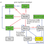
Bubonic plague switches between two forms during its life cycle: A) biofilm-forming, less-virulent form and B) non-biofilm-forming, virulent form.
The plague germ adopts biofilm-forming, less-virulent form in low ambient temperature (21 degrees Celsius) such as flea digestive tract. The biofilm blocks the flea’s gut and induces the flea to bite more frequently, increasing disease transmission. A master regulator gene RovM promotes biofilm formation and represses RovA. Virulence factors are downregulated so that the germ doesn’t kill the flea immediately.
The plague germs adopt highly virulent form under high ambient temperature (37 degrees Celsius) such as inside body of warm-blooded animal. RovM is repressed under high temperature. RovA becomes activated and promotes production of virulence factors.
I don’t want to explain too explicitly. You are welcome to read these papers. Basically, Gen. Yang tweaked with bacterial signaling pathway to create super lethal mutant plague germs that produce virulence factors consistently.
References:
1. Chen H, Cao S, Zhou Y, Wang T, Jiao Y, Tan Y, Wu Y, Ren Y, Song Y, Zhang JR, Du Z, Yang R. Molecular turn in Yersinia pestis pathogenesis: implications of the gppAframeshift for bacterial survival in human macrophage. Emerg Microbes Infect. 2025 Dec;14(1):2467778. doi: 10.1080/22221751.2025.2467778. Epub 2025 Mar 3. PMID: 39945756; PMCID: PMC11878169.
pubmed.ncbi.nlm.nih.gov/3994…
2. Liu L, Fang H, Yang H, Zhang Y, Han Y, Zhou D, Yang R. Reciprocal regulation of Yersinia pestis biofilm formation and virulence by RovM and RovA. Open Biol. 2016 Mar;6(3):150198. doi: 10.1098/rsob.150198. PMID: 26984293; PMCID: PMC4821237.
pubmed.ncbi.nlm.nih.gov/2698…
3. Liu Z, Gao X, Wang H, Fang H, Yan Y, Liu L, Chen R, Zhou D, Yang R, Han Y. Plasmid pPCP1-derived sRNA HmsA promotes biofilm formation of Yersinia pestis. BMC Microbiol. 2016 Aug 4;16(1):176. doi: 10.1186/s12866-016-0793-5. PMID: 27492011; PMCID: PMC4973556.
pubmed.ncbi.nlm.nih.gov/2749…
4. Gao X, Wang M, Liu Z, Bi Y, Song Y, Yang R, Han Y. Altered Yersinia pestisvirulence is associated with the small regulatory RNA HmsA encoded on the plasmid pPCP1. Future Microbiol. 2020 Sep;15:1207-1215. doi: 10.2217/fmb-2019-0319. PMID: 33026884.
pubmed.ncbi.nlm.nih.gov/3302…
Also read the paper below. A CCP scientist and US scientists collaborated on Yersinia research (just comparative genetics, not GOF). “This research was supported by the Intramural Research Program of the NIH, NIAID.”
Chouikha I, Sturdevant DE, Jarrett C, Sun YC, Hinnebusch BJ. Differential Gene Expression Patterns of Yersinia pestis and Yersinia pseudotuberculosis during Infection and Biofilm Formation in the Flea Digestive Tract. mSystems. 2019 Feb 19;4(1):e00217-18. doi: 10.1128/mSystems.00217-18. PMID: 30801031; PMCID: PMC6381227.
pubmed.ncbi.nlm.nih.gov/3080…


