

Pathologist who loves a good diagnostic puzzle 🔬 | Educator at heart | Here to share cases, tips & the occasional plot twist under the microscope
Joined March 2020
- Tweets236
- Following868
- Followers936
- Likes968



Anna Vasileva retweeted

Hard time remembering all those #hemepath immunostains/markers? This chart by Dr. Jeanette Ramos will help! Full chart free here: kikoxp.com/posts/20554.
#medstudents #pathTwitter #path2path #usmle #MedStudentTwitter #MedED #medtwitter #hematology #hemeonc #lymphoma #leukemia

Anna Vasileva retweeted

1/3 #GIpath #CritterOnTwitter Look what I found in this inflamed antral hyperplastic gastric polyp 🤓. At first glance, I thought that it is hallucinations or illusions due to end of the week fatigue 😂 then I started to take images of every single one. So all are represented 😅


Anna Vasileva retweeted

#PathQuiz 🔬🦋 #ThyroidPath
A) NIFTP
B) High-grade follicular cell-derived thyroid carcinoma
C) Oncocytic Carcinoma
D) Invasive Encapsulated Follicular Variant of Papillary Thyroid Carcinoma
#Pathology #ThyroidPath

ALT A 48-year-old woman presents with a solitary, well-demarcated thyroid mass. Capsular invasion. @IARCWHO

ALT Note the unequivocal penetration through the thick fibrous capsule and into adjacent vascular spaces.
Anna Vasileva retweeted

ADH in Radial Scar 🫚🔬
Occasionally, atypia can be spotted within sclerosing lesions—here, a monotonous epithelial proliferation partially filling ducts within a fibroelastotic, architecturally distorted background.
#PathX #PathTwitter #breastpath

Anna Vasileva retweeted

Progestin effects in benign endometrium
What you see:
- Surface papillations
- Dilated glands
- Breakdown, necrosis, and plasma cells
Dr. Castrillon #USCAP2026 #pathology #PathX #PathTwitter #everydayGYN

Anna Vasileva retweeted

Patient with classic papillary thyroid carcinoma in both lobes, here is a lymph node from the central compartment. What do you make out of it?
Short thread follows:

Anna Vasileva retweeted

Counting eos is not enough 🔍
15 eos/hpf supports EoE, but EoE-HSS captures true disease activity by scoring 8 histologic features.
See for yourself: cchmc.pathpresenter.net/publ…
#PediPath #GIpath #EoE #MedEd


Anna Vasileva retweeted

Illustration of a case of Follicular Lymphoma with Hyaline Vascular Unicentric Castleman Disease-"like" changes➡️
🩸Depleted germinal centers (Fig 2)
🩸Onion-skinning of mantle zones (Fig 2-3)
🩸Penetrating hyalinized vessels, imparting a lollipop-like configuration of germinal centers (Fig 3)
🩸Twinning (fusion) of follicles (Fig 4)
This case turned out to be postive for BCL2 translocation by FISH, with an aberrant CD10-positive population by flow cytometry analysis.
More on this variant of Follicular Lymphoma➡️pubmed.ncbi.nlm.nih.gov/2887…
To complicate things, the "follicular variant" of Hyaline Vascular Unicentric Castleman Disease, with characteristic back-to-back follicular architecture, may greatly mimic follicular lymphoma as well ➡️pubmed.ncbi.nlm.nih.gov/8116…
➡️pubmed.ncbi.nlm.nih.gov/1903…
In summary, by morphologic examination, Follicular Lymphoma may present like Hyaline Vascular Unicentric Castleman Disease; Conversly, Hyaline Vascular Unicentric Castleman Disease may show a presentation very similar to Follicular Lymphoma
#hemepath #lymsm #PathX #PathTwitter #MedX #MedTwitter #MedEd


Anna Vasileva retweeted

Breast, Fibrocystic Changes, Multiples Intraductal Papillomas, Gross pics, #pathology, #pathologists


Anna Vasileva retweeted

For our 2023 #pathology trainees, remember this #GUPath adage👇
“Not all renal tumors with clear cells are clear cell renal cell carcinoma”
… spot on in this molecular era!
👇Below are DDX for Clear cell #RCC including #renal tumors with some degree of cytoplasmic clearing🔬

Anna Vasileva retweeted

Tenosynovial Giant cell tumor
Arises from synovium (joints; bursae; tendon sheath)
Due to CSF1 mutation
Localized type usually on the fingers
Treatment of localized: surgical excision
#pathtwitter #pathsig #orthotwitter #medtwitter #MedStudentTwitter #softtissue


Anna Vasileva retweeted

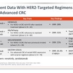
Targeted therapies for colorectal cancer have advanced greatly since my last review on this subject in 2016 with the incomparable @JLHornick. MMR deficiency, high TMB, BRAF, HER2 and numerous rearrangements have all become targetable.

Anna Vasileva retweeted

Classic example of collagenous micronodules (aka mucinous fibroplasia) in #prostatecancer #GUPath #pathologists @Pathologists


Anna Vasileva retweeted
E-cadherin expression in invasive or in-situ carcinoma DOES NOT preclude classification as lobular
Dr, Harrison #USCAP2023 #everydaybreast #breastpath #pathology #pathtwitter #pathologist #pathology #pathresidents

Anna Vasileva retweeted
Bladder
Histological subtypes of urothelial carcinoma and those with divergent differentiation are all considered high-grade tumors-WHO 5th edition
Dr. Paner USCAP23 #USCAP2023 #PathTwitter #GUpath #pathology #pathologist #pathresident #pathfellow

Anna Vasileva retweeted

One in a million Monday.
Unwittingly saved by the clinician. This case was not a consult, it was just a good old biopsy from a satellite hospital for whose patients we provide pathology diagnostic services. In that hospital, breast surgeons coordinate breast patient care, perform biopsies, discuss treatment and surgical plans with patients, and eventually refer to oncology as needed. Unlike our radiologists and oncologists here at WashU, the surgeons at that hospital do not want to get a tumor diagnosis then get an addendum with the biomarkers. They like to see a complete report all at once. I can't say I dislike that practice, and after this particular case, I like it even more, because it saved me from a major (albeit exceptionally rare) misdiagnosis.
The images below depict a tumor I was squarely going to call invasive solid papillary carcinoma because, to my eyes, that's exactly what it looks like. Except that's not what it is. And because I waited for the biomarkers to sign it out, I was fortunate to find out it was triple negative, which would be extremely unusual for solid papillary carcinoma. So, I contacted the surgeon and dug into her chart, and sure enough, the patient had a history of metastatic mucoepidermoid carcinoma of the minor salivary glands. That's when I started seeing a hint of squamous differentiation and possible mucous cells. CK5/6 and p63 were done and were confirmatory. MAML2 FISH was also positive and sealed the deal. I did not find a similar case in the literature.
Lessons from this monumental near miss.
1. Clinician preferences did save me this time, but the clinician's omission of pertinent medical history is a common and persistent problem.
2. Always remember that breast biomarkers are not only predicitve and prognostic, but also diagnostic. If their pattern does not fit with the tumor's morphology, take a deep breath and dive into those muddy diagnostic waters.
3. Again and again, be vigilant, look for clinical information, don't make assumptions. Easier said than done, of course, and I do not want to encourage paranoia because I am not the paranoid type, but sharp alertness is one absolute must, and not allowing hasty explanations for things that don't quite fit is another.
4. Morphology is key, but nature loves mimicry. Don't be a hammer. Everything is not a nail.
@washupathedu @wusm_pathology #PathTwitter #Breastpath #ENTpath

ALT Uncanny resemblance to invasive solid papillary carcinoma

ALT Hints of squamous differentiation with intercellular bridges and orangeophilic tadpoles, and a lonesome possible mucous cell

ALT p63

ALT CK5/6
Anna Vasileva retweeted
High yield 💎appendiceal lesions.
- Difference between diverticula and LAMN
#surgpath #pathology #PathTwitter #GIpath
Check it online 📱digital.olivesoftware.com/ol…
Thanks for sharing @Voltaggiol 🌟


This Post is from an account that no longer exists. Learn more
Anna Vasileva retweeted
Ovary
Sertoli-Leydig cell tumor
Grade of differentiation
-Tubular differentiation of the Sertoli cell component (decreasing with increasing grade)
-Quantity of primitive gonadal stroma (increasing with increasing grade)
-Leydig cells also decrease with increasing grade
WHO and DAPA GR #EverydayGYN #gynpath #PathTwitter #pathologists #pathresidents #pathfellow

Anna Vasileva retweeted
Inflammatory Breast Conditions
Central and Peripheral
Dr. Mais #USCAP23 #breastpath #everydaybreast #pathology #PathResident #pathfellow #PathTwitter #pathologists


Anna Vasileva retweeted

There are prominent granular cell features in this IDHwt GBM with morphology much akin to macrophages (largely negative for CD68). These have been referred to as granular cell astrocytoma or granular cell GBM.
Goes to show how variable the morphology can be in #glioblastoma.
